Understanding and Managing a Popped Calf Muscle

Comprehending the makeup of our body, particularly in areas susceptible to frequent injuries, is the first step towards its adequate care, and the prevention or remediation of any potential harm. The calf muscle, one of the most commonly injured areas, plays a critical role in our daily activities, allowing us to walk, run, and jump. But, when this muscle pops or gets injured, it can result in significant pain and discomfort. This knowledge will delve into the intricacies of the calf muscle’s anatomy, the common causes and symptoms of a popped calf muscle, potential diagnoses and treatments, and some effective preventive measures to mitigate the risk of such injuries.

1. Anatomy of the Calf Muscle

Anatomy of the Calf Muscle

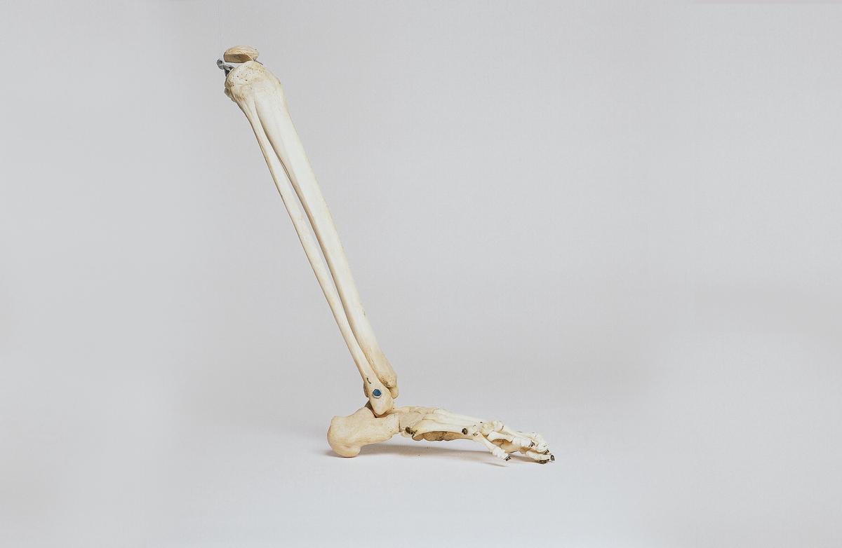
The human anatomy consists of two significant muscles forming what we commonly refer to as the calf muscle: the gastrocnemius muscle and the soleus muscle. These muscles, located at the back portion of the lower leg, play a crucial role in human movement, particularly in walking, running, and jumping.

The larger and more visible of the two is the gastrocnemius muscle. This muscle has two parts or “heads,” which give the calf its distinctive shape. It starts behind the knee on the thighbone, stretches down the back of the leg, and attaches to the heel bone with the Achilles tendon’s help.

Just underneath the gastrocnemius is the soleus muscle. Unlike its counterpart, the soleus is a broader, flatter muscle that provides stability and maintains posture. Its function mainly revolves around actions like standing or walking, where we keep our legs straight. It also attaches to the heel bone alongside the gastrocnemius through the common tendon, the Achilles.

Working together, these muscles help push the foot down – a movement called plantar flexion. They can contract powerfully to make the body move forward or upward. Additionally, the calf muscles help pump blood back up from the legs to the heart – a critical aspect of the circulatory system.

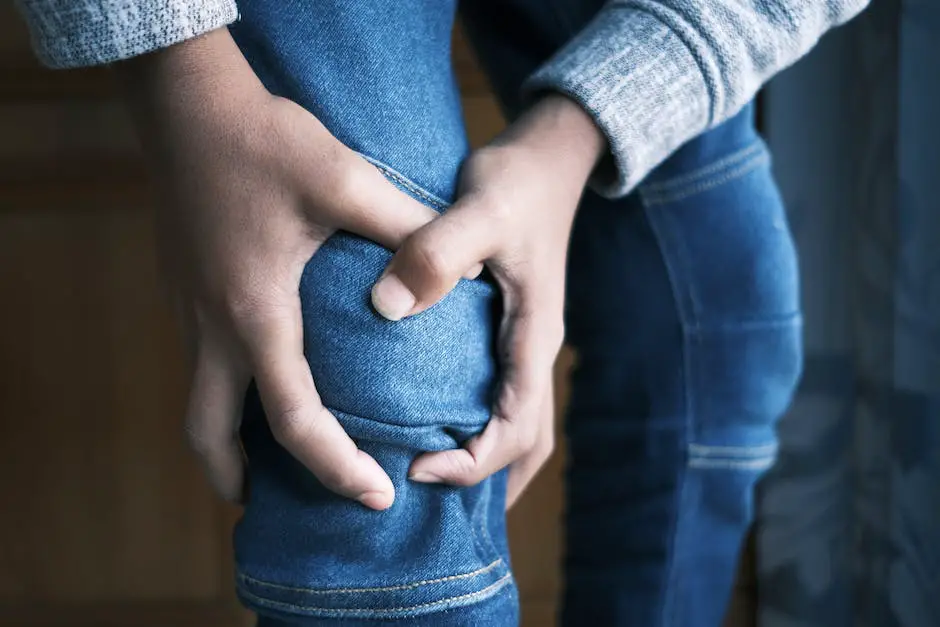
Pop in the Calf Muscle and Pain

A sudden pop in the calf muscle, often associated with intense pain, could signify a common injury known as a calf strain or “pulled” muscle. This can occur when the muscle fibers in the calf are stretched beyond their limit, resulting in damage or tears to the small fibers.

The ‘pop’ feeling users commonly report might be moderate (grade 1), causing minor damage to few muscle fibers and resulting in some pain and discomfort, severe (grade 2), where a larger amount of fibers are damaged, causing more substantial pain and difficulty moving, or worst-case (grade 3), a complete rupture of the calf muscle.

For minor to moderate strains, symptoms may include a sharp pain or immediate soreness during activity, swelling, and difficulty walking. For more severe injuries, you might notice a visible bruise or depression where the tear occurred, sensitivity to touch, or even an inability to stand or put weight on the injured leg.

When a pop occurs, immediate treatment involves rest, ice, compression, and elevation (RICE). By managing the injury promptly, you can help alleviate pain, decrease swelling, and protect the injured calf muscle. Be sure not to strain the muscle further, avoid heat, and never attempt to stretch or massage an acutely strained muscle.

Introduction to Calf Muscles

The calf muscles are inherent to the functioning of our musculoskeletal system, significantly contributing to our locomotive abilities and the flow of blood in our bodies. A comprehensive understanding of their anatomical structure, physiological role, and the potential indicators of injury can help preserve their health and smooth operation.

Illustration of the anatomy of the calf muscle, showing the gastrocnemius and soleus muscles, along with their attachments and functions.

2. Causes and Symptoms of a Popped Calf Muscle

Understanding the Phenomenon of Calf Muscle Pop

The popping of a calf muscle, technically referred to as a calf strain or tear, transpires when these muscles undergo excessive stretching. This overextension could cause the muscle tissues to rupture, ensuing a popping sensation.

Certain activities known to possibly induce a calf muscle pop encompass explosive actions like sprinting, jumping, or any leg-intensive exertion, and sudden directional changes in sports. Undergoing these activities without an optimum warm-up could escalate the risk of calf injury as the unprepped muscles might not withstand the stress.

Apart from strenuous physical activities, underlying health complications can also lead to a calf muscle pop. Prior injuries in the area, especially those inadequately rehabilitated, can predispose the muscles to future tears. Leg or foot structural abnormalities, such as flat feet, can exert irregular pressure on the calf muscles, leading to a rupture. Lastly, the aging process itself poses a risk as it generally results in diminished muscle elasticity and strength, thereby making them more injury-prone.

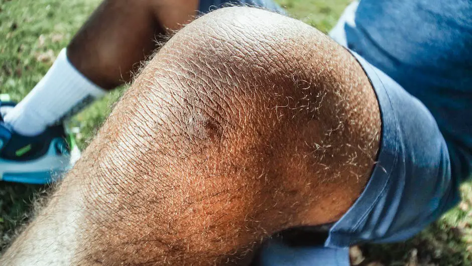
Identifying the Symptoms of a Popped Calf Muscle

When a calf muscle pops, the symptoms might present immediately and intensely, or they could manifest more slowly and subtly, all depending on how serious the tear is.

One of the most typical symptoms is pain, which can vary in intensity from a dull ache to a piercing, sharp sting. This discomfort usually amplifies when the person stands or walks and can further intensify with activities that involve straining the calf muscles, such as running or jumping.

Another tell-tale symptom of a torn muscle is a sensation of popping or snapping at the time of the injury, usually accompanied by a sharp, sudden pain in the calf.

Other potential signs include swelling or bruising, difficulties in walking or flexing the foot particularly on inclined or uneven surfaces, diminished mobility in the ankle joint. Some may even experience muscle spasms or cramps. In severe instances, a palpable gap, dent, or visible abnormality where the muscle has torn might be noticeable.

Should you suspect a popped calf muscle, it is crucial to consult with a healthcare professional to prevent further damage and promptly initiate the required treatment.

Image depicting the causes of a popped calf muscle, including physical activities, previous injuries, structural abnormalities, and age-related muscle weakness.

3. Diagnosis and Treatment Options

Detecting a Popped Calf Muscle: the Diagnostic Process

The evaluation of a popped calf muscle often commences with a physical check-up carried out by a medical professional. While conducting the physical examination, the doctor may apply slight pressure to the affected region to spot any situational swelling or deformation, and to measure the severity of the pain. Functional exercises that require calf muscle movement might also be put forward to test your agility.

Complementary to the physical examination, imaging tests such as X-rays, ultrasound, and MRI scans often serve as additional diagnostic tools. An X-ray can reveal a more grievous injury like a fracture, and ultrasound and MRI scans provide comprehensive imagery of the soft tissues, which aids in spotting muscle tears or strains.

Aiming to eliminate the likelihood of deep vein thrombosis (DVT), which can portray symptoms similar to those of a popped calf muscle, doctors might ask for an ultrasound or a D-dimer blood test. D-dimer is a specific protein that appears in the blood only after a blood clot has broken down, hence its increase can suggest the presence of DVT.

Addressing a Popped Calf Muscle

Dealing with a popped calf muscle effectively requires understanding the extent of the injury and the overall health condition of the individual. The intervention options are usually determined by these factors.

The R.I.C.E strategy is widely recommended as the initial remedial measure for minor to moderate muscle injuries. This approach encompasses Rest (staying away from activities that cause pain), Ice (application of ice packs on the injured region for 20-minute intervals to minimize swelling), Compression (using an elastic bandage to stop further swelling and offer support), and Elevation (raising the leg above heart level when feasible to decrease swelling).

To alleviate pain and minimize inflammation, over-the-counter analgesics such as Ibuprofen may be suggested. Engaging in certain stretching exercises could also promote recovery.

In cases of severe muscle tear, intervention from a medical expert might be necessary. This could involve a surgical repair of the muscle, followed by immobilization with a cast or splint to foster healing. Post this, a round of physical therapy may be required to regain muscle strength and encourage flexibility.

The exact treatment strategy could vary based on specific situations. Some possibilities include prescription of muscle relaxants or recommendations for platelet-rich plasma (PRP) injections to expedite recovery. PRP therapy involves drawing the patient’s blood, treating it to increase platelet concentration, and injecting it back into the injured area to amplify the body’s innate healing capabilities.

Given the individual nature of each situation, it’s crucial to adhere to the directions and treatment plan prescribed by a healthcare professional for an efficient and suitable treatment.

Illustration of a person holding their calf muscle with an arrow pointing to the affected area

4. Effective Preventive Measures for Calf Muscle Injury

Proactive Steps to Prevent Calf Muscle Injuries

Injuries to the calf muscle often occur during sports and other physical activities. The sensation or sound of a ‘pop’ in the calf muscle usually indicates a strain or tear, which may cause immediate pain and discomfort. However, there are several proactive steps one can take to mitigate the risk of such injuries.

Targeted Exercises

Targeted exercises focusing on the calf muscles not only strengthen this area of the body but also make it more resilient to injury. There are several exercises one can incorporate into their routine. Calf raises, for instance, can enhance both endurance and strength. This exercise involves standing upright and lifting your heels off the ground, engaging the calf muscles. Incorporating stretches, such as the towel stretch or standing calf Stretch, can also improve flexibility and reduce rigidity, which, in turn, lowers the risk of muscle strains or tears.

Correct Warm-ups and Cooldowns

Warming up before engaging in any strenuous physical activity is vital. It increases blood flow to the muscles, loosens joints, and can prepare the body for the demands of exercise. An effective warm-up often includes light cardio activity, such as jogging or cycling, and dynamic stretching that mimics the movement of the upcoming activity. Similarly, cooling down allows a gradual reduction in heart rate and helps flush metabolic waste from muscles. Gentle stretches and slow, controlled movements can minimize post-activity stiffness and aid recovery.

Proper Hydration

Staying hydrated before, during, and after exercise can work wonders for preventing muscle injuries. Dehydration can cause muscle cramps, which may lead to injury. Therefore, drinking plenty of fluids helps maintain electrolyte balance, supporting muscle function and potentially preventing muscle damage.

Identifying Warning Signs

Understanding the body’s warning signs can aid in injury prevention. If you experience persistent muscle tightness, fatigue, or sharp pain, it may be a timely indication to take a break or modify your routine. Feeling as though a muscle has “popped” or “snapped” could be a sign of a calf muscle tear, requiring immediate cessation of the activity and prompt medical attention.

To sum up

Consistent strength training and stretching exercises, paired with thorough warm-ups and cool-downs, are crucial elements in the battle against calf muscle injuries. In addition, staying well-hydrated and listening to your body’s signals can significantly reduce the risk of experiencing pain or a pop in the calf muscles.

Illustration showing calf muscles and preventive measures for calf muscle injury

Knowledge about the anatomy of the calf muscle, the potential causes of injuries, and the corresponding symptoms are vital for early detection and treatment. Understanding the diagnosis process and potential treatment options can help you make informed decisions if faced with such a scenario. Implementing effective preventive measures, including targeted exercises, proper hydration, correct warm-up, and cooldown routines, can greatly reduce the risk of enduring a popped calf muscle. Be in tune with your body, recognize the warning signs, and take timely action to maintain your body’s fitness and health.

Mastering the Knee Pop Pose: A Simple Guide

The world of yoga is vast and diverse, with a myriad of poses designed to cleanse the mind, strengthen the body, and facilitate a deeper connection with oneself. One such structure, the knee pop pose, is the focus of this in-depth analysis. With nuances in its origin and various benefits, understanding the knee pop pose surpasses mere physical practice. Circumscribing within its realm are fundamental components such as its definition, origin, purpose, and the physiological benefits it delivers. Additionally, recognizing the required physical preparation and the meticulous execution further enhances the significance of the knee pop pose within the broader yogic discipline. This exploration serves to empower you in confidently and safely engaging in this pose, amplifying its benefits, and coming to respect its role in personal fitness and wellness.

Understanding the Knee Pop Pose

Understanding the Knee Pop Pose

The knee pop pose, also known as the “Popping Asana”, is a familiar sight in the realm of Yoga, distinctive for its peculiar position where a yogi pops their knee, meaning extending it forcibly. The pose involves a gentle twist of the spine while one leg is straight and the other is bent with foot on the other thigh or knee.

Origins of the Knee Pop Pose

Rooted in the ancient art of Yoga, the origins of the knee pop pose can be traced back to traditional Indian practices. Yoga, as a unifying discipline, seeks to balance the mind, body, and spirit through various physical and meditative techniques. The unique knee pop pose is one such technique designed to instill balance and harmony in one’s mental and physical state. The pose earned its name because of the ‘popping’ or ‘snapping’ sensation felt in the knee during the position, which indicates a comprehensive stretch.

Purpose of the Knee Pop Pose within Yoga

The knee pop pose serves several purposes in Yoga. First and foremost, it helps stretch the lower body, more specifically the hips, thighs, and knee joints. Furthermore, it works on stabilizing the spinal column. The pose is known to aid in strengthening the lower body, enhancing body awareness, and improving balance. It is an one-legged posture that combines a sense of grounding through the standing leg with an openness expressed through the hip of the lifted leg.

Benefits of the Knee Pop Pose

When performed correctly, the knee pop pose brings a host of benefits to the practitioner. It not only improves balance and stability but it also stretches and strengthens the muscles on the hips, hamstrings, quadriceps and the lower back. These factors contribute to an overall enhanced physical fitness. On a more profound level, the pose can have therapeutic effects, potentially alleviating sciatica, menstrual discomfort, and digestive problems. By promoting better body alignment and posture, it can also alleviate backaches and stiffness often associated with sedentary lifestyles.

Understanding the knee pop pose, its origins, implications within yoga, and the benefits it brings to the body when properly executed, lays the groundwork for effectively learning and integrating this pose into your yoga routine. With sufficient practice, the knee pop pose can be a brilliant addition to your repertoire, enhancing your flexibility, strength, and overall well-being.

Illustration of a person performing the knee pop pose, with one leg straight and the other bent with foot on the other thigh or knee.

Physical Preparation for the Knee Pop Pose

Physical Preparation

Before attempting the knee pop pose, it’s vital to begin with a warm-up routine to increase your body’s temperature and make your muscles more pliable. You can start with some gentle aerobic activities such as brisk walking or jogging. This will help get the blood flowing through your body and can prevent injury during the pose.

Following the aerobic warm-up, it’s time to stretch your muscles and joints, particularly the ones that will be in use during the knee pop pose. These include your knees, thighs and hips. Some recommended stretches could include lunges, leg swings, quad stretches, and hip openers. Spend at least 5-10 minutes on this part of your warm-up, gradually deepening your stretches as your body becomes more flexible.

Strength and Flexibility Training

Before moving to the actual knee pop pose, it is vital to strengthen and stretch specific muscle groups. The thighs, hips and knees need to be strong and flexible to successfully perform the pose without injury. Some exercises that can help prepare your body include squats, lunges, and leg curls for strengthening, and lunges, butterfly stretches, and hip flexor stretches for flexibility.

Additionally, incorporating yoga into your routine can help improve overall flexibility and strength. Positions like the downward dog, warrior pose, and tree pose can particularly benefit the muscle groups involved in the knee pop pose.

Technique Practice

In the days or weeks leading up to attempting the knee pop pose, be sure to practice lower body poses and balancing techniques. This will help your body prepare for the balance and coordination required for the knee pop pose. If you’re practicing yoga, consider adding poses like the chair pose or the extended side angle pose to your routine.

Cooling Down

After practicing these preparatory exercises or the knee pop pose itself, it’s crucial not to neglect your cool down routine. This is as important as the initial warm-up. It allows your body to recover from the vigorous activity and helps prevent muscle stiffness and soreness. Gentle stretches can help reduce muscle tension and promote relaxation. Drink plenty of water to replenish any fluids lost during your workout and consider using a foam roller to massage any particularly sore or tense muscles.

Remember, it takes time and consistent effort to master any new physical skill, including the knee pop pose. Listen to your body and avoid rushing the process to prevent injuries. If you experience any persistent pain or discomfort, it is advisable to seek professional guidance.

Illustration showing a person performing the knee pop pose, demonstrating the stretching and balance required.

Executing the Knee Pop Pose

Body Alignment in the Knee Pop Pose

The first step to correctly executing the knee pop pose is to ensure that you are properly aligned. Stand tall with your feet spread hip-width apart. Your body should be straight, from your head down to your feet. The key is to maintain even weight distribution between both your feet.

Positioning Your Feet

Next, slightly rotate your feet so they are turned inwards and pivot on the heels. In doing so, your toes should be pointing towards each other. Once achieved, push your knees forward while maintaining your feet firmly planted on the floor. This movement will cause your knees to pop outward.

Popping the Knees

Keep your body erect and gently “pop” or push your left knee backwards. Following that, repeat the process with your right knee, keeping in sync with the beat of your chosen music. Start with slow, deliberate knee movements until you are comfortable executing them, then improve speed and rhythm as practice progresses.

Modifications for Beginners

If you are a beginner, you can modify the knee pop pose to make it easier. One way to do this is by placing your hands on your thighs for support while executing the ‘pop’. Another modification is to perform the pose near a wall, using it for balance and support.

Potential Mistakes to Avoid

There are common mistakes experienced beginners should seek to avoid when doing the knee pop pose. These include bending the upper body forward, uneven weight distribution, and making the popping movement too large. Also, in your enthusiasm to execute the pose, remember to avoid forcing your knees out of their natural alignment. This can potentially lead to knee injuries.

Observing Correct Body Alignment

Keep a constant awareness of your body alignment while practicing the knee pop pose. This includes checking that your feet are properly positioned, your weight is evenly distributed, and your upper body is perpendicular to the floor. The knee pop pose requires coordination and attention to ensure it’s done correctly and safely.

Practice under Professional Supervision

For beginners, it is always recommended to practice the knee pop pose under the guidance of a professional instructor. They will supervise your movements, ensure proper technique, and provide instant feedback and corrections. This way, you can successfully learn the pose and significantly reduce the risk of potential injuries.

Illustration showing the correct body alignment and foot positioning for the knee pop pose

Residing at the crossroads of fitness and mindfulness, the knee pop pose embodies a myriad of benefits. Physically, it strengthens and extends various body parts and encourages appropriate body alignment, fostering overall fitness. Philosophically, learning this pose is equally about embracing a discipline that dates back centuries, honouring its roots. The journey through understanding, preparation, and execution of the knee pop pose is a venture of self-engagement that transcends beyond a physical accomplishment. It is about carving out a more robust, healthier version of oneself, gaining profound insights into one’s physical and mental prowess. Once armed with an understanding and appreciation of this pose, it is an enlightening doorway to delve deeper into the enriching world of yoga.

Understanding a Torn Calf Muscle: Causes, Symptoms, and Treatments

Calf muscles are an integral part of our daily mobility, supporting movements such as walking, running, or jumping. Understanding the nuances of a torn calf muscle is crucial, whether you’re an athlete, an active person, or you have experienced sudden discomfort in your lower leg. Through this comprehensive examination, we dive deep into the anatomy of a torn calf muscle, investigate the common causes, discuss the evident symptoms, and explore diagnosis methods, treatment options, and subsequently prevention and rehabilitation strategies. Armed with this information, you’ll be better prepared to identify and manage a torn calf muscle should you ever face this predicament.

What is a Torn Calf Muscle?

Understanding a Torn Calf Muscle

A torn calf muscle, also known as a calf strain, is a common sports injury that occurs when the muscle fibers in the calf are overstrained or stretched beyond their limits. The calf is composed of two muscles, the gastrocnemius and the soleus, which join at the Achilles tendon to attach at the heel. When either of these muscles is overstretched, the muscle fibers tear, resulting in a muscle strain.

Grades of a Calf Muscle Tear

Calf muscle tears are typically categorized into three different grades, depending on the severity of the injury:

  • Grade 1: This level involves minor muscle tear where less than 10% of the muscle fibers are damaged. The pain is often mild and you may be able to continue physical activity with minimal discomfort.
  • Grade 2: This moderate muscle tear involves damage to a more substantial amount of muscle fibers, often accompanied by an acute, sharp pain and inability to continue physical activity.
  • Grade 3: This is a severe muscle tear where the muscle is completely ruptured or torn. This injury usually causes severe pain, inability to walk, and significant bruising.

Identifying the Symptoms of a Torn Calf Muscle

A torn calf muscle is commonly characterized by a sudden, sharp pain in the back of the leg. Quite often, this intense pain is accompanied by a snapping or popping sensation which signifies the actual occurrence of the tear. The muscle might tighten immediately, and this acute pain might gradually transform into a throbbing ache that is aggravated by movements or attempts to use the injured leg.

Post injury, it is typical to have difficulty standing on toes or flexing the foot downwards. In extreme cases, this may result in the individual adopting a limp or, in severe situations, becoming incapable of walking due to unbearable pain and pressure.

Body’s natural response to an injury, such as swelling and bruising, is typically seen with a calf muscle tear due to an increased flow of blood to the damaged region. The skin around the torn muscle may feel warm due to heighted circulation aiming at muscle repair.

While a torn calf muscle can cause severe discomfort and can immobilize an individual temporarily, remember that with adequate rest and care, such injuries usually heal. Yet, medical intervention becomes necessary if the pain is extreme, if walking becomes impossible or if recurring calf muscle strains are experienced.

Illustration showing calf muscle injury with dashes instead of spaces

Causes of a Torn Calf Muscle

Understanding the Causes of a Torn Calf Muscle

A torn calf muscle or calf strain can occur to anyone and is typically seen in individuals who engage in athletic activities, fail to stretch properly before exercises or make sudden, intense movements.

Sports activities, especially that involve running and jumping, or other dynamic actions, are a key source of calf muscle tears. Athletes engaged in sports such as basketball, tennis, football, or even regular runners, are particularly susceptible to such injuries. During theses activities, the calf muscles undergo constant contraction and relaxation and any abrupt change or increased force can result in a tear.

The absence of proper muscle warm-up or stretching before physical activities too can lead to injuries like a torn calf muscle. Without adequate stretch or warm-up, muscles remain less flexible and cold, thereby being more prone to injury. Consequently, stretching is crucial to prepare the muscles for any physical exertion. Without stretching, muscles might get abruptly yanked or torn as they are forced into high-intensity activity.

Torn calf muscles are also often a result of sudden intense movements commonly seen in sports and other physical activities, like abrupt changes in direction, acceleration or deceleration, which all put substantial strain on the calf muscle. This might push the muscle to stretch beyond its limits and result in a tear.

While understanding the common causes can help avoid such painful injuries, it is important to know that despite taking precautions, sometimes injuries do occur.

Understanding a Torn Calf Muscle and Its Symptoms

Curious to know what a torn calf muscle feels like? The sensation can differ significantly based on the extent of the injury, however, it typically starts off with a sudden, intense pain. Many individuals describe feeling a “pop” or a sharp, stinging sensation in the lower part of the leg, often around the ankle. The injured area may quickly become swollen, tender, and start to take on a reddish hue. In severe instances, standing or walking may prove difficult or even impossible. Over time, the injured spot may start to show signs of bruising.

When the calf muscle is affected by strain or tear, many experience the discomfort at the back of the lower leg, mainly when the foot is flexed or during walking. The sensations may range from a minor twinge, which might be felt only during strenuous activities, to unbearable pain which makes everyday tasks like standing or walking a challenge. The intensity of pain and the certainty with which it can be attributed to a torn calf muscle is a good indication of whether medical attention should be sought out.

In essence, a torn calf muscle can result in significant discomfort and incapacitation. Prompt therapeutic intervention is crucial. A doctor or a physiotherapist can assess the extent of the tear and prescribe appropriate treatment, which may involve rest, application of cold packs, compression dressings, elevation of the affected leg, analgesics, and physiotherapy sessions.

An image showing a person holding the back of their lower leg in pain.

Symptoms and Diagnosis of a Torn Calf Muscle

Early Symptoms Following a Torn Calf Muscle

The most immediate symptoms of a torn calf muscle often involve a sudden popping or snapping sensation in the back of your leg. This is often accompanied by intense pain which might disrupt the activity you were engaged in. Some people even compare this searing pain to a gunshot or an impact on the leg.

Right after the injurious incident, you might find it challenging to bear weight on the affected leg, thereby limiting your mobility. You may face difficulty in walking or even just standing still. Swelling generally manifests in the lower leg and in some instances, bruising might also be observed if blood vessels have been affected during the injury. You might also notice muscle weakness or complete inability to use the calf muscle, a sign which is termed as muscle paralysis.

Progression of Symptoms Over Time

As time passes, the acute pain may subside a bit; however, you will continue to feel discomfort, especially when engaging in activities involving the calf muscle such as standing on tiptoe or walking uphill. Some people may also experience muscle spasms, a tightness in the back of the lower leg, and tenderness when touching the calf.

The swelling gradually increases over a few hours to a few days and can spread to your ankle and foot. The bruising might change colors from blue and black initially to yellow or green as it starts to heal.

Examining a Torn Calf Muscle

If you’re experiencing persistent pain, swelling, and other signs, it’s highly recommended to seek the advice of a healthcare professional. A physical examination is likely to be conducted. During this assessment, the doctor will carefully press and manipulate the affected area to determine whether there are any abnormalities.

Whenever the symptoms signal a severe tear or rupture, further imaging tests may be scheduled. Ultrasound, which creates detailed images by reflecting sound waves off body tissues, can show the calf muscle and show the damage. Alternatively, Magnetic Resonance Imaging (MRI) can be used, which employs a strong magnetic field and radio waves to deliver comprehensive images of your body’s interior.

Under rare and complex circumstances, doctors might suggest an arthroscopy. This minimally invasive procedure is used to gain a clear study of the injury by inserting a tiny camera into the joint via a small incision.

Conclusively, a torn calf muscle is represented through various symptoms that might seem intense initially and may alter over time. The precise health evaluation and imaging tests help establish the seriousness of the injury and suitable treatment methods.

Illustration of a torn calf muscle showing the affected area

Treatment Options for a Torn Calf Muscle

Getting to Know a Torn Calf Muscle

A tear in the calf muscle is typically the result of overstraining or overextending this lower leg muscle. Athletes frequently experience such injuries, particularly those participating in high-impact sports or activities that require sudden speeding up or slowing down. The presence of a torn calf muscle is usually marked by a sharp, electric-like pain in the back of the calf, often likened to being hit from behind.

Such sharp pain often coincides with a “popping” feeling, with pain levels varying from sharp or throbbing to dull or achy. The pain usually intensifies with strenuous activity, especially any action requiring push-off from the injured leg. In severe cases, the pain may even hinder walking.

Pain is not the only symptom of a torn calf muscle, other indicators include swelling, bruising, muscle weakness, and a restricted lower leg movement range. The injured calf can show signs such as reddening and feeling warm to touch, depending on the injury’s severity.

Non-Surgical Treatments for Torn Calf Muscle

Non-surgical treatment methods are often the first line of remedy for a torn calf muscle. The most common methodology is the RICE protocol: rest, ice, compression, and elevation. By avoiding activities that aggravate the pain, applying ice to minimize swelling, wearing a compression bandage to support the muscle, and elevating the leg, individuals often experience pain reduction and healing.

Physical therapy is another common non-surgical treatment. It encompasses exercises and stretching routines designed to restore flexibility and strength in the calf muscle. Therapy may start with light stretching, gradually progressing to more strenuous activities as the muscle heals. The goal is to restore the muscle’s normal function and to prevent future injury.

Pain management is also key in treating a torn calf muscle. Over-the-counter pain relievers such as ibuprofen or acetaminophen may be effective. In more severe cases, a physician might prescribe stronger prescription medications.

Treatment and Recovery Following Calf Muscle Tear Surgery

When a tear in the calf muscle is severe or non-surgical treatments can’t render the desired effects, surgery may be the only solution. This generally involves an orthopedic surgeon cutting away the damaged muscle and stitching the healthy tissue back together.

Post-operative care might require the lower leg to be immobilized with a cast or brace for a few weeks to facilitate the repair process. Just like with non-surgical care, physiotherapy is a key part of rehabilitation after surgery. A bespoke routine of carefully calibrated stretching and strengthening exercises can significantly accelerate recovery.

Variations in recovery time hinge on the degree of injury and the overall health status of the individual patient. While minor calf strains can take just a few weeks to heal, a serious muscle tear requiring surgical intervention might need several months for complete recovery.

Illustration of a torn calf muscle with dashed lines indicating the tear and surrounding areas of redness and swelling

Prevention and Rehabilitation

Identifying the Symptoms of a Torn Calf Muscle

Commonly referred to as a calf muscle strain, a torn calf muscle can trigger a range of symptoms that span from a sudden intense pain to a nagging, dull ache. The rear of the lower leg might feel stiff and tender, and the pain could intensify with physical activity. Swelling and bruising are other likely signs, with the severity aligning with the extent of the injury. Some individuals liken the sensation to a powerful hit or a ‘pop’, succeeded by escalating pain. Actions like standing on the toes or walking might amplify the discomfort. Other indications of a torn calf muscle are a limited range of motion, inflammation, and loss of muscle strength.

Prevention Measures for a Torn Calf Muscle

Preventing further injury to a torn calf muscle involves avoidance of activities that may exacerbate the pain or cause more harm. This can include jumping, running, or other intense physical activities without proper warmup.

Keeping the calf muscles strong can also serve as a preventative measure. Regular exercises such as calf raises, leg presses, and heel drops help to reinforce the muscle and improve overall strength.

Stretching before and after workouts is also essential in maintaining muscle flexibility, which can help, mitigate the risk of injuries. It’s recommended to perform dynamic stretches before training and static ones afterward to facilitate muscle recovery, flexibility, and mobility.

Dietary Changes to Support Calf Muscle Health

An often-overlooked aspect of muscle health is diet. Consuming optimal levels of protein help in muscle recovery and growth since they serve as the primary building blocks for muscle tissue. Adding good sources of carbohydrates and healthy fats can also fuel recovery.

Adequate hydration should never be neglected either. Water plays a critical role in most processes in the body, including muscle contraction and nutrient transport.

Rehab Exercises for a Torn Calf Muscle

After the initial healing phase, rehab exercises are crucial in regaining the strength you previously had in your calf muscle. These exercises should start gently and increase in intensity as the healing progresses.

One of the simplest exercises to start with is calf pumping. While seated, simply pump your foot up and down, like pressing a pedal. This will gently work the calf muscle and increase blood flow to the area, promoting healing.

Progress to weight-bearing exercises such as calf raises when comfort levels allow. Stand on a flat surface and raise your heels off the ground, standing on your toes, before lowering back down.

Lastl,y stretching can aid in the healing process. Calves are often tight muscles, so gentle stretches can help increase flexibility and strength. A simple calf stretch can be done by facing a wall and pressing the injured foot back, keeping the heel on the ground and the leg straight.

Remember, healing times can vary and rushing through rehab can lead to more damage. It is always beneficial to seek professional advice to ensure the exercises are appropriate for your specific condition and stage of recovery.

Illustration depicting a torn calf muscle injury, showing the calf muscle with redness, swelling, and a tear.

Understanding injuries like a torn calf muscle is an essential step towards effective treatment, recovery, and the prevention of further damage. Emphasizing the importance of knowing the causes, symptoms, and treatment options cannot be overstated in helping speed up recovery and reducing discomfort. Moreover, it is pivotal to follow preventative measures and rehabilitation process keenly to regain full functionality and avoid reinjury. Staying informed is a significant part of maintaining our health and wellness, allowing us to lead an active and fulfilling lifestyle. Here’s to your health and a long life filled with mobility and strength!

Effective Arthritis in Knees Remedy and You

The increasing prevalence of arthritis, a condition marked by inflammation and pain in joints, is a critical health issue affecting millions worldwide. This discourse aims to provide a thorough understanding of arthritis in the knee, one of the most widely affected regions, delving into its causes, symptoms, types, and effects. It offers an enlightening exploration into non-surgical treatment options, which includes a study of pain relievers, anti-inflammatory drugs, corticosteroids, physical therapy, and the role of weight management, braces, and assistive devices. Furthermore, it highlights the surgical remedies available for knee arthritis, their potential pros and cons, recovery process and expected outcomes. It also helps to shed light on a myriad of natural therapies and home remedies used by patients and their efficacies.

Understanding Arthritis

Understanding Arthritis in the Knees

Arthritis in the knees is a joint condition characterized by the inflammation and degeneration of the knee joint. The resulting loss of normal joint structure and function leads to pain, stiffness, and reduced mobility. There are several types of arthritis that can affect your knees, each with unique causes and consequences.

Arthritis Types

The most common type of arthritis in the knees is osteoarthritis. This is a degenerative joint disease that typically occurs in individuals 50 years and older. It manifests as the cartilage that acts as a cushion in the joints is worn away, leading to bones rubbing together and causing pain.

Rheumatoid arthritis is another type of arthritis that affects the knees. It is an autoimmune disorder where your body’s immune system attacks the lining of your joints, leading to discomfort and swelling. Post-traumatic arthritis is a form of osteoarthritis that develops after an injury to the knee, such as a ligament tear or bone fracture.

Causes of Knee Arthritis

Developing arthritis in the knees happens over time due to wear and tear on the joints. This degeneration is often the result of aging, but certain factors can accelerate the process. Obesity, for instance, increases pressure on the knee joints and can lead to faster wear of the cartilage. Injuries to the knee can also trigger early onset of arthritis. Lastly, genetics plays a role, as those with a family history are more likely to develop the condition.

Symptoms and Effects of Knee Arthritis

The primary symptoms of arthritis in the knee are pain and stiffness in the joint. As the condition progresses, these symptoms can become more persistent and severe, eventually affecting the individual’s ability to comfortably perform everyday activities. The loss of stable joint structure can result in a decreased range of motion, and the degree of pain experienced can range from mild to severe, often worsening after physical activity.

Complications and Risk Factors

Complications caused by knee arthritis can include chronic pain, deterioration of joint tissues, and decreased mobility. In severe cases, arthritis can lead to deformity of the joint and disability. Risk factors that increase the likelihood of developing knee arthritis include obesity, age, a history of knee injury, and certain occupations that require repetitive stress on the knee joint.

In summary, arthritis in the knees is a common condition that affects many people as they age. By understanding its causes, symptoms, and effects, you can better manage the condition and maintain your joint health.

Illustration depicting the effects of arthritis in the knees, showing inflamed joints and limited mobility.

Non-Surgical Treatment

Non-Surgical Remedies for Knee Arthritis

Arthritis in the knee is a common ailment that causes pain, swelling, and reduced mobility. Fortunately, several non-surgical remedies exist to manage the symptoms of this condition effectively.

Pain Relievers and Anti-inflammatory Drugs

Over-the-counter pain relievers, including acetaminophen (Tylenol), ibuprofen (Advil, Motrin), and naproxen sodium (Aleve), can help to decrease pain and inflammation caused by knee arthritis. These drugs can have potential side effects such as liver damage, kidney problems, or gastrointestinal distress, so they should be taken as directed by your healthcare provider.

Corticosteroids

Corticosteroids, such as prednisone or cortisone, are another treatment option for knee arthritis. These drugs can be taken orally or injected directly into your knee by your healthcare provider. Corticosteroids work by reducing inflammation and decreasing the body’s immune response. While effective, long-term usage can lead to weakening of tissues and potential infections.

Physical Therapy

Physical therapy, including exercises that enhance strength, flexibility, and range of motion, can be highly beneficial for patients with knee arthritis. Working with a physical therapist who specializes in knee conditions can help you customize a program that works best for your body and increases your mobility without causing more pain.

Impact of Weight Loss

Carrying extra weight puts more stress on your knee joints, which can speed up the progression of arthritis. Hence, losing weight, if overweight, can significantly improve your symptoms. By changing your diet to healthier options and physically active lifestyle will not only help in weight loss but also improve your overall health.

Braces and Assistive Devices

Braces can be worn to support your knees and help distribute weight more evenly. This can reduce pain and inflammation. Assistive devices, such as canes or walkers, can also provide support and improve your mobility if your knee condition is severe.

Understanding Your Treatment

It’s important to understand that treatment effectiveness may vary from person to person, and what works best for one person may not work as well for another. Always consult with your healthcare provider before starting a new treatment, understand potential side effects, and monitor your body’s response to treatment. This can help you and your healthcare provider determine the next step in your treatment plan, ensuring you get the best care for your knee arthritis.

Image depicting different treatment options for knee arthritis, including braces, physical therapy, and pain relievers.

Surgical Treatments

Arthroscopy for Knee Arthritis

Arthroscopy is a minimally invasive surgical procedure on a joint in which an examination and sometimes treatment of damage is performed using an arthroscope. For knee arthritis, the surgery involves the insertion of the arthroscope, a small camera, into the knee joint, providing a clear view of the inside of the knee. This enables the surgeon to investigate the severity of your arthritis and possibly treat the issue at the same time.

The pros of arthroscopy are its relatively quick recovery time (usually four to six weeks), the small surgical scars and the lower risk of complications compared to more invasive surgeries. The cons are that it may not provide long-term relief if your arthritis is severe or widespread.

Joint-Preserving Osteotomy Surgery

Osteotomy is a surgical procedure that involves cutting and reshaping a bone near a damaged joint. This surgery can realign the knee, shifting weight from the damaged area to a healthier part of the knee. This procedure can be suitable if arthritis affects primarily just one side of your knee.

On the upside, osteotomy can significantly reduce pain, prolong the lifespan of the knee joint, and delay the need for knee replacement surgery. Drawbacks include a relatively long recovery period of several months and potential complications like discomfort and infection.

Joint Replacement Surgery for Knee Arthritis

Joint replacement surgery, also known as arthroplasty, involves removing the damaged joint surfaces and replacing them with artificial parts. This surgical procedure can be a total knee replacement (TKR) or a partial knee replacement (PKR).

A notable advantage of joint replacement surgery is the significant reduction in pain and improvement in quality of life. Many people regain mobility and can return to their regular activities. However, cons include a long recovery time, usually between three and six months, possible complications such as infection, and the potential need for revision surgery if the artificial joint wears out.

In conclusion, surgical treatments for knee arthritis have their distinct advantages and disadvantages. Decision-making should involve discussion with your doctor about your overall health, the extent of your arthritis, and your lifestyle and expectations. Remember that surgical treatment is typically considered only after non-surgical methods have failed to provide satisfactory relief.

Illustration of a surgeon performing knee arthritis surgery

Photo by nci on Unsplash

Natural Therapies

Acupuncture as Arthritis Remedy

Acupuncture is a traditional Chinese therapy that practitioners use to balance the flow of energy or life force — known as qi or chi (CHEE) within the body. The technique involves inserting thin needles into specific points in the body. It is believed to stimulate nerves, muscles, and connective tissue to improve blood flow and boost the body’s natural painkillers. Some scientific studies suggest that acupuncture works particularly well on people with osteoarthritis in the knees. While acupuncture is generally considered safe when performed by an experienced practitioner, some potential side effects include soreness, minor bleeding or bruising where the needles were inserted.

Hydrotherapy for Arthritis Relief

Hydrotherapy involves the use of water, both hot and cold, for health purposes. For arthritis in the knees, hydrotherapy exercises in a warm-water pool can help to decrease pain and improve physical functioning. Water provides a low-impact, therapeutic environment that can help reduce the load on your knees, allowing you to perform exercises that might otherwise be painful or difficult. A regular hydrotherapy routine can provide lasting benefits for individuals with osteoarthritis. However, it’s essential to note that therapy should be carried out under supervision for safety purposes.

Diet Modification for Arthritis Management

Diet plays a significant role in managing arthritis. Foods rich in antioxidants, such as fruits and vegetables, can fight inflammation and strengthen the immune system. Meanwhile, omega-3 fatty acids, found in fish like salmon and mackerel, can also help reduce symptoms. Limiting processed foods and adding more whole grains to your diet can also help balance your body’s inflammation response. Each person’s body responds differently to dietary changes, so it’s recommended to keep a food diary to monitor the impact of diet on arthritis symptoms.

Arthritis-Targeted Exercise Routines

Exercise is one of the best ways to combat arthritis in the knee. Low-impact activities like walking, cycling, and swimming can increase your range of motion, improve joint flexibility, and strengthen the muscles around your joints, thus providing added support. Strength-building exercises like weightlifting can also help by supporting the joint and reducing stress on it. While exercise is beneficial, it’s crucial to start slow and gradually increase intensity to avoid injury. It’s recommended to work with a physical therapist or trained professional to develop an effective and safe exercise routine.

Using Natural Supplements for Arthritis

Natural supplements like glucosamine and chondroitin, turmeric, and omega-3 fatty acids can help reduce arthritis symptoms. Glucosamine and chondroitin, which are components of normal cartilage, are commonly used for treating osteoarthritis. Turmeric’s active compound curcumin has strong anti-inflammatory effects. Fish oil supplements, rich in omega-3 fatty acids, have also been shown to reduce joint pain and stiffness. However, supplements can interact with other medications, so it’s important to talk to a health provider before starting any new supplement regimen.

While these natural therapies and home remedies can aid in managing arthritis symptoms, it’s essential to consult with a healthcare provider for personal recommendations based on individual health status.

A person receiving acupuncture treatment for arthritis

Knee arthritis, despite being a pervasive condition, can be effectively managed with an arsenal of treatments and therapies. Evaluating non-surgical treatments, surgical options as well as natural remedies, it’s possible to reduce the symptoms, improve joint health and enhance the quality of life for the affected individuals. The therapeutic approach can be tailor-made to suit every individual’s unique condition and lifestyle. By familiarizing ourselves with each aspect of this condition, we can navigate the path to better knee health and overall wellbeing. Hence, we can be empowered to make informed decisions that will ultimately serve the best personal outcomes.

Natural Solutions for Curing Diabetes

Diabetes, a chronic condition affecting millions globally, poses significant health challenges. But despite the perceived complexity, captivating success stories and a growing body of scientific evidence suggest that simple yet powerful lifestyle modifications and natural remedies may be key in managing or even reversing this disease. From understanding the intricate physiology of diabetes to spotlighting natural treatments and the significance of regular medical check-ups, this exploration sheds light on how individuals can actively participate in their healthcare journey and potentially combat diabetes naturally.

Understanding Diabetes

Understanding Diabetes: A General Overview

Diabetes is a chronic health condition that affects how your body turns food into energy. There are different types of diabetes including: type 1, where the body does not make insulin; type 2, where the body does not use insulin well; and gestational, which develops in some women during pregnancy and usually goes away after birth. Each type differs and carries its unique characteristics.

Insulin is a hormone made by the pancreas, and it lets the glucose from the food you eat enter your cells to be used as energy. In cases where the body either can’t produce insulin or is resistant to it, glucose stays in the bloodstream which leads to high blood sugar levels.

Causes of Diabetes

The factors causing diabetes differ for each type. Type 1 diabetes has been linked to genetics and possibly certain viral infections, while type 2 diabetes is caused by genetics and lifestyle factors such as obesity and lack of physical activity. Gestational diabetes may be caused by hormonal changes or genetics.

How Diabetes Affects the Body

In the short term, people with diabetes may experience excessive thirst and urination, fatigue, blurred vision, slow healing wounds, and more. Long term effects can be even more severe. High blood sugar levels can damage blood vessels and nerves, leading to heart disease, kidney disease, stroke, and even blindness.

Symptoms and Complications of Diabetes

The symptom of diabetes can vary depending on the sugar level in your body. They may include frequent urination, extreme thirst or hunger, fatigue, blurry vision, cuts/bruises that heal slowly, weight loss even when eating more (type 1 diabetes), tingling, pain, or numbness in hands or feet (type 2 diabetes).

Left unregulated, diabetes can lead to more serious complications. It can damage the blood vessels, impacting the heart, brain, and legs. It can also cause neuropathy (nerve damage), nephropathy (kidney disease), problems with the skin and eyes, and gum infections.

The Importance of Managing Diabetes

It’s crucial to regularly monitor and manage your blood sugar levels if diagnosed with diabetes. This can prevent complications and promote a healthier life. Regular exercise, eating a balanced diet low in the sugar and high in fruits, vegetables, and whole grains, not smoking, consuming alcohol in moderation, and maintaining a healthy weight can all aid in managing and even preventing the progression of diabetes.

Nature’s Approach to Managing Diabetes

No concrete cure exists for diabetes as of now, but incorporating natural solutions can provide support in managing this health condition. Indulging in physical activity on a regular basis can result in lower blood glucose levels and an increase in insulin sensitivity. Adopting a nutritious diet that comprises leafy vegetables, lean sources of protein, and complex carbohydrates can help keep blood sugar levels in check. In addition to this, integrating herbal supplements like cinnamon, fenugreek, and aloe vera can show promising results in terms of managing blood sugar levels.

However, it’s imperative to engage with a health care professional prior to beginning any new therapeutic regimen. Guidelines suggested by them could steer you towards a safe and efficacious journey towards diabetes management.

Image showing a person with a glucometer and healthy food choices, representing diabetes management

Lifestyle and Dietary Changes

Shaping Lifestyle and Diet for Improved Health

A key aspect of effectively managing, and possibly naturally neutralizing diabetes, evolves from alterations in lifestyle. These changes consist of a blend of dietary modifications, amplification in physical activities, strategies for weight reduction, stress management techniques, enhanced sleep patterns, and maintaining proper hydration.

Dietary Choices

A healthy diet plays a significant role in blood sugar control. Consuming a whole food, plant-based diet rich in fruits, vegetables, whole grains, lean proteins and healthy fats can help reduce blood sugar levels. The American Diabetes Association suggests focusing on foods with a low glycemic load, as they prevent blood sugar spikes. Regular, balanced meals are also encouraged. Portion management is vital; overeating, even healthy food, can raise blood sugar levels.

Physical Activity

Exercise is another essential component in managing diabetes naturally. Regular physical activity helps regulate insulin sensitivity and supports healthy weight management, both of which are critical for blood sugar control. Activities such as walking, jogging, swimming or cycling for at least 150 minutes per week can have a considerable impact. It is advisable to include resistance training as part of workout regimen twice per week.

Weight Management

A healthy body weight can significantly improve insulin sensitivity, decreasing the body’s need to produce insulin. Weight loss, especially around the midsection, can help prevent and reverse type 2 diabetes. Small, consistent changes in diet and exercise may lead to sustainable weight loss and control.

Stress Management

Stress hormones can lead to increased blood glucose levels. Regular exercise, deep-breathing exercises, yoga, meditation, counseling, and support groups can help maintain and manage stress levels, thereby controlling blood sugar levels.

Sleep Quality and Quantity

Experts have found links between inadequate sleep and increased risk of diabetes. Poor or insufficient sleep impairs the body’s ability to use insulin effectively resulting in higher blood glucose levels. Thus, improving sleep quality and ensuring sufficient sleep of 7-9 hours per night can be useful in diabetes management.

Hydration

Staying well-hydrated aids in controlling blood sugar levels. Besides water, other beverages like herbal teas can be beneficial. Sweetened drinks, however, may cause blood sugar spikes and should be avoided.Attention to research underscores how managing diabetes naturally through lifestyle alterations is not just a myth, but a reality. Carrying out these strategies requires determination and a high degree of self-discipline, but the benefits can be significant. These include reduced dependency on medication, decreased diabetes symptoms, and an overall enhancement in your quality of life.
Image description: A person holding a plate with fruits, vegetables, and healthy food, representing a healthy lifestyle and diet changes.

Natural Remedies

Natural Supplements and Herbs for Diabetes Care

A range of herbs and supplements are frequently recommended as aids in diabetes management. These generally work by either improving digestion or affecting insulin sensitivity. For instance, cinnamon is a supplement that has shown promise in increasing insulin sensitivity and lowering blood sugar levels. On the same note, fenugreek seeds have the ability to slow down the breakdown of carbohydrates, resulting in reduced blood sugar levels.

That said, although these supplements showcase positive outcomes, it’s vital to understand that they cannot supersede standard insulin treatments. Instead, they should be viewed and used as a supportive therapy in tandem with prescribed diabetes medication. Before including any new herbs or supplements into your diabetes care routine, it’s highly advisable to speak with a healthcare professional.

The Role of Yoga in Managing Diabetes

Yoga, a practice combining mental, physical, and spiritual disciplines, carries numerous potential benefits for individuals with diabetes. Regular yoga practice can enhance glucose metabolism, decrease insulin resistance, and promote weight loss, which are all beneficial in managing blood glucose levels. Additionally, yoga can potentially help manage stress and mental well-being, which is often important for individuals struggling with chronic illnesses.

Specific yoga postures, often referred to as asanas, can stimulate pancreas function and control blood sugar levels. For instance, poses like Vajrasana (Diamond Pose) and Mandukasana (Frog Pose) stimulate the functioning of the pancreas, making them beneficial for diabetics. However, it is important to learn these postures from a trained instructor to ensure appropriate form and prevent injury.

Acupuncture for Diabetes

Acupuncture is a traditional Chinese medicine practice wherein fine needles are inserted into specific points across the body. According to some small-scale studies, acupuncture has shown potential in managing diabetes symptoms and complications. This includes improving glycemic control, increasing insulin sensitivity, and alleviating diabetic neuropathy pain.

Within acupuncture, there are specific points targeted for blood sugar control such as Pishu (BL20) and Weishang (BL21), which are said to boost pancreatic function. However, more large-scale, rigorous research is needed to establish acupuncture as a standard care practice for diabetes.

Understanding Potential Risks and Precautions

Undeniably, natural remedies for diabetes can show beneficial effects, but alongside come certain precautions and potential side effects. It is important to realize that some supplements and herbs may not react favorably with conventional diabetes medications, possibly perpetuating or altering their respective impacts.

In addition, natural physical activities such as yoga can certainly aid management. However, proper technique is of utmost importance to prevent injuries. In cases of acupuncture, a certified professional should always be involved in order to adhere to safety standards.

To summarize, it’s vital to grasp that these natural remedies for diabetes management are ideally used as supplements to traditional diabetes care rather than substitutes. It is highly encouraged to always seek advice from a healthcare provider before implementing any significant alterations to your treatment regimen.

Illustration of different herbs and supplements bundled together with a diabetes medical symbol.

Role of Regular Medical Check-ups

The Crucial Role of Regular Health Check-ups in Diabetes Management

Regular health check-ups are invaluable in the ongoing management of diabetes. They gain even more importance when combined with natural health practices. As a chronic health issue, diabetes demands consistent and rigorous management to ward off complications and maintain wholesome health. Natural practices like a tailored diet, regular exercise, and supplements can keep a check on blood sugar levels as well as support the health of the pancreas. However, periodic medical check-ups enhance this approach by securing the overall well-being of individuals with diabetes.

Routine Screening and Management of Diabetes

Routine screening is essential in the management of diabetes. Regular blood glucose tests, for instance, can help monitor the effectiveness of your lifestyle and dietary choices in controlling diabetes. If your blood sugar levels are not within the target range, your healthcare provider can recommend changes to diet, exercise regimen, or medication.

Apart from blood glucose tests, regular check-ups might also involve foot and eye examinations, blood pressure checks, kidney function tests, and cholesterol level evaluations. These screenings help detect any diabetes-related complications early, allowing for timely intervention.

Routine screenings also provide the opportunity to review and adjust treatment plans. This could involve a change in medication type or dosage, introduction of supplementary nutritional plans and natural remedies, or modifications to exercise routines.

Types of Tests to Monitor Diabetes

Several different tests are used in monitoring diabetes. The most common is the glycated hemoglobin (A1C) test, which measures your average blood sugar level over the past 2 to 3 months. Your healthcare provider may use this test to diagnose diabetes and prediabetes.

Regular self-monitoring of blood glucose at home using a blood glucose meter or continuous glucose monitoring (CGM) device helps track daily blood sugar levels and understand their relationship with factors like food, physical activity, and stress.

Other routine tests include regular urine tests to check kidney function, lipid profile tests to monitor cholesterol and triglyceride levels, and annual retinal screenings to detect diabetic retinopathy.

Complementing Natural Approaches with Medical Assistance

While natural approaches can be extremely beneficial, they should be used alongside regular medical check-ups and not as a standalone treatment. Using natural remedies like maintaining a balanced diet, keeping active, and nutritional supplementation supports self-managed care. However, the technical expertise of a healthcare professional is crucial in providing a holistic, well-rounded approach to diabetes management.

Healthcare professionals offer immediate corrections and adjustments to treatment plans based on regular screenings. They also provide the much-needed support and education on living with diabetes, which is essential in building the confidence and skills you need to manage the condition effectively.

It’s essential to understand that regular medical check-ups complement natural approaches in managing diabetes. These routine visits allow for early detection and timely intervention, consistent analysis and modification of treatment strategies, and continual health education and support, all of which contributes to effective diabetes management.

Image depicting a person visiting a doctor for a check-up

Success Stories and Case Studies

Thomas C’s Journey to Natural Recovery

Life presented Thomas with a significant challenge when he was diagnosed with Type 2 diabetes at the age of 45. Faced with constantly fluctuating blood sugar levels and the prospect of lifelong medication, Thomas felt overwhelmed. His curiosity piqued by an online community advocating natural remedies and lifestyle alterations, Thomas saw a glimmer of hope and decided to explore this approach. He made a significant U-turn in his lifestyle, incorporating plant-based nutrition, a disciplined exercise regimen, and daily mindfulness meditation. Astonishingly, Thomas began to notice a steady decline in his blood sugar levels. Six months into his new lifestyle, Thomas had lost 30 lbs, with improved health to an extent that his doctor took him off medication.

Belinda’s Victory Over Diabetes

Belinda has been a type 1 diabetic since the age of 16. Her story is one of true grit and determination. Belinda had always been passionate about fitness, so when she was diagnosed, she resolved not to let this condition control her life. Along with an insulin regimen, she integrated a strict workout routine complemented by a carefully monitored diet into her daily life. Belinda also started attending a support group that practiced yoga and holistic self-care. These changes brought about a significant improvement in her blood sugar control. Fifteen years after her diagnosis, Belinda’s doctor announced that she had effectively “reversed” her diabetes and was showcasing the health parameters of a non-diabetic.

John Beats the Odds

John was diagnosed with gestational diabetes during his wife’s third pregnancy. A simple blood test revealed that his fasting blood sugar levels were dangerously high. Instead of being prescribed medication, John’s doctor suggested making lifestyle changes first. With the help of a dietician, John started eating smaller portions, consuming a balanced diet, and drastically reducing his intake of sugary and starchy foods. He also started to exercise regularly, beginning with gentle walks that gradually evolved into jogging and weightlifting sessions. After a year, John had lost a dramatic 50lbs and was no longer classified as diabetic. His doctor was amazed at the powers of lifestyle modification.

Sarah Finds Balance

Sarah faced an uphill battle with uncontrolled diabetes for almost 10 years. She struggled with frequent episodes of hypoglycemia and hyperglycemia. Reading about herbal remedies sparked her interest, and she started digging deeper. She learnt about the Chinese herb, Berberine, and it’s supposed benefits in blood sugar regulation. After consulting with a naturopathic doctor, who confirmed its safety, Sarah started taking Berberine supplements, and her blood sugar control improved significantly. This natural treatment, along with a balanced diet and exercise, helped Sarah reduce her insulin dosage substantially, finding a better balance in her life.

Image of people celebrating their successful management and reversal of diabetes.

Throughout this exciting exploration, we’ve unpacked the wealth of possibilities in natural diabetes management – from dietary changes, weight management, and stress management to natural remedies and the necessity of regular health check-ups. Each individual’s journey with diabetes is unique, and these strategies may serve as stepping stones towards a healthier future. In the mosaic of diabetes care, the narratives of real people who have effectively reversed their diabetes remind us all that, while challenging, a more natural approach to diabetes future holds immense promise and potential.

Understanding and Alleviating Soleus Tightness

The soleus muscle, tucked within the mighty muscles of your calf, plays a significant, albeit often-underestimated, role in our daily mobility. Its function extends far beyond enabling movement and bears a directly causal relationship with your overall health and quality of life. Yet, much like many unsung heroes of our physiology, the soleus muscle tends to get sidelined, leading to tightness. This tightness, while seemingly insignificant, can have lasting impacts on our well-being. This exploration seeks to shed light on all aspects of soleus tightness, from its causes and symptoms to its treatment and prevention and, importantly, its impact on one’s quality of life.

Understanding the Soleus Muscle

The Soleus Muscle: Anatomy & Function

The soleus is a powerful muscle located in the calf, deep to the more noticeable gastrocnemius muscle. It extends from just below the knee to the heel and along with the gastrocnemius, forms the Achilles tendon. The function of the soleus muscle involves plantarflexion of the foot, which refers to the movement that points the foot downward, like when you push the gas pedal in your car.

The soleus muscle plays a critical role in movement and stability. It enables us to stand upright, walk, run, and even jump. Moreover, it is crucial in maintaining balance and posture. Importantly, the soleus muscle also aids in pumping blood back to the heart from the lower body due to its role in the calf muscle pump.

Understanding Soleus Neglect and Tightness

Despite its importance to overall mobility, the soleus muscle is often neglected in regular strength training and stretching routines. This neglect can lead to muscle tightness. Tightness in the soleus muscle often presents as a deep, achy feeling at the back of the lower leg. It may become apparent when walking, running or standing for extended periods. In severe cases, it could even impair mobility.

Soleus muscle tightness can arise due to various reasons. It is common among athletes, particularly those in sports that require repetitive running or jumping. Moreover, sedentary lifestyle or jobs requiring prolonged sitting or standing can also contribute to soleus muscle tightness. Poor footwear and inappropriate training routines, especially neglecting to stretch before and after exercise, also play a role.

Soleus Tightness and Mobility

Soleus tightness can lead to various complications if not addressed promptly. As part of the Achilles tendon, tightness in this muscle can put additional pressure on the tendon, leading to conditions such as Achilles tendonitis. It can also negatively impact mobility, affecting gait and balance, which could present a risk for falls or other muscle strain.

Furthermore, tightness in the soleus muscle may lead to a condition called compartment syndrome, characterized by pressure build-up in the muscle, leading to pain and potential injury.

Consistent workout routines, including stretching and strengthening exercises, can effectively prevent the soleus muscle from tightening. Utilizing massage therapy and tools like foam rollers can also aid in lessening muscle tightness. However, should a tight soleus impair mobility to a significant degree or result in enduring pain, it is crucial to seek professional medical intervention to avoid further complications.

Illustration of the soleus muscle, showing its location in the calf and its connection to the Achilles tendon.

Causes of Soleus Tightness

Getting to Know the Soleus Muscle

Located at the back of the lower leg (the calf), the soleus is a robust muscle integral to movements such as standing, walking, and running. The role it plays involves flexing the ankle joint and stabilizing the ankle and lower leg, absorbing shock, and generating propulsion while walking or running in conjunction with the other muscles in the lower leg and foot. Notwithstanding, a tight soleus muscle can lead to a range of issues, including discomfort and notable reductions in performance levels and mobility.

Common Causes of Soleus Tightness

There are several causes of soleus tightness, each affecting individuals in different ways. Among the primary culprits is overuse, or too much stress on the muscle from high-impact or strenuous activities such as long-distance running, jumping sports, or even prolonged standing. Overuse can cause the muscle fibers to become shortened, leading to tightness and discomfort.

Improper stretching is another leading cause of soleus tightness. Without regular, proper stretching, the muscles can become stiff and less flexible, making them more prone to injury. This includes not just the soleus, but also other muscles that work in conjunction with it.

Anatomical imbalances, such as differences in leg length, foot arch, or muscle strength can create undue stress on the soleus, leading to tightness. For example, an individual with flat feet may overstrain their soleus muscle because it has to work harder to maintain balance and stability.

Other medical conditions can also lead to tightness in the soleus. For instance, conditions that cause inflammation such as tendinitis or compartment syndrome can result in swelling and tightness in the muscles around the affected area. Metabolic conditions such as diabetes or peripheral vascular disease may also affect the elasticity and functionality of the muscle, leading to tightness.

Effects of Activities and Lifestyles

Activities and lifestyles can have a profound impact on soleus health. Sedentary lifestyles involving long periods of sitting may lead to soleus tightness as lack of movement can cause muscles to shorten and stiffen. High-impact sports and activities can overwork the muscle, causing it to become strained and tight. Even daily repetitive activities like wearing high heels or climbing stairs can contribute to a tight soleus muscle, due to constant and repetitive straining.

Conversely, activities promoting flexibility, strength, and balance, such as yoga, pilates, and targeted strengthening exercises, can protect against soleus tightness and other muscle-related discomforts. It is essential to incorporate regular stretching, particularly post-activity stretching, to maintain flexibility and muscle health.

Research and Prevention of Soleus Tightness

It’s important to note that while soleus tightness can impact anyone, certain factors may put you at higher risk. These include unavoidable factors like age, working in professions requiring prolonged standing or repetitive movements, high levels of physical activity, or past experience with muscle injuries. There are also a few preventive strategies that can be embraced to counteract the possible risk, including hydrating properly, consuming a balanced diet, engaging in routine stretching and strengthening exercises, and wearing suitable footwear.

Illustration depicting the soleus muscle and its role in ankle flexion and stability

Symptoms and Diagnosis of Soleus Tightness

Identifying Symptoms of Soleus Tightness

Soleus tightness is generally recognizable by a notable decrease in flexibility, particularly when trying to stretch the calf muscles, which leads to discomfort or difficulty. The sensation of this tightness can be described as a constant feeling of fatigue or heaviness in your calves – a sensation that becomes more pronounced after being on your feet for a while.

Additionally, the presenting symptoms might include a persistent dull pain that appears to travel from your calf muscle down towards your ankle. Physical activities like walking, running, or jumping might magnify this pain.

In some cases, the discomfort stemming from the tight soleus muscle may extend to the Achilles tendon since this muscle is directly connected to the heel bone. This happens because the strain from the tightness inflames this crucial tendon and may result in cases of extreme inflammation or, worse still, a total or partial rupture of the tendon.

Complications of Soleus Tightness

Left untreated or unattended, soleus tightness could lead to more severe complications. Persistent discomfort or pain in the feet can upset an individual’s overall gait and posture, leading to additional problems like lower back or hip pain. In athletes and physically active individuals, soleus tightness can significantly limit performance by reducing their speed and power.

Understanding the Diagnosis of Soleus Tightness

Soleus tightness is typically identified in a medical examination that takes into account your history, any previous injuries or surgeries, and your lifestyle habits that may be contributing to the problem. This comprehensive evaluation helps physicians get a clear picture of the potential causes of your discomfort.

The physical examination forms a crucial part of this process. It involves assessing your flexibility, muscle strength, and overall range of motion to gauge the extent of soleus tightness. A variety of simple exercises may be involved, such as standing on tiptoes or flexing the foot with a straightened leg in a bid to induce any possible discomfort or limitations.

If necessary, your physician may further recommend imaging tests like X-rays, MRIs, or ultrasounds for a detailed view of your leg structures. These tests aid the medical professionals in ruling out any other potential causes of discomfort, as well as pinpointing any signs of damage or inflammation in the muscle tissues or tendons involved.

Based on these findings, clinical interventions may be recommended to relieve discomfort, improve muscle flexibility, and prevent complications. This might include physical therapy exercises, regular stretching routines, and even lifestyle modifications to reduce strain. If pain persists or is severe, more invasive treatments, such as injections or surgery, may come into play.

Illustration depicting symptoms and complications of soleus tightness.

Treatment and Prevention of Soleus Tightness

Defining and Identifying Causes of Soleus Tightness

Soleus Tightness can be described as an uncomfortable feeling of stiffness or strain in the soleus muscle. This muscle is an integral part of your calf running from just below the knee to the heel. It’s crucial to various movements, including walking, running, and balance maintenance. Often, overuse of this muscle, inadequate stretching before and after exercise, and a sedentary lifestyle can contribute to its tightening or inflammation.

Common Symptoms of Soleus Tightness

Symptoms of soleus tightness may include pain or discomfort in the calf when walking or running, tenderness to the touch, decreased motion range, and weakness in the calf muscle. Chronic soleus tightness can limit mobility and lead to other issues such as plantar fasciitis or Achilles tendinitis if left untreated.

Treatment Methods for Soleus Tightness

If you’re suffering from soleus tightness, treating it early can save you from potential complications. At-home treatments for soleus tightness often involve self-massage, heat, cold, and stretching. Regular application of a heating pad can enhance blood circulation and promote healing. On the other hand, cold packs can reduce inflammation and relieve pain. Self-massage using foam rollers or massage balls can also alleviate muscle tension.

In many cases, incorporating specific stretching exercises into your routine can be beneficial. Exercises that specifically target the soleus muscle include seated and standing calf raises, stair raises, towel stretch, and yoga poses like downward-facing dog and warrior I. It’s advisable to perform these exercises under the guidance of a physical therapist or a certified trainer to prevent injury.

In severe cases, where at-home remedies do not suffice, professional medical intervention may be required. Treatment options can range from physical therapy sessions to surgery in extreme cases.

How to Prevent Soleus Tightness

The best way to stave off soleus tightness is through proactive preventative measures. Incorporated into your daily routine they can maintain and improve the flexibility of your calf muscles. Regular stretching exercises before and after physical activities are especially beneficial. A balanced diet and proper hydration contribute to overall muscle health, including that of the calf muscles. Shoes that specifically offer arch support can help reduce strain on the calves, and regular low-intensity exercises such as walking or swimming strengthen and maintain calf flexibility.

Being aware of potential warning signs is equally vital. Constant or recurring pain, discomfort, or even swelling in the calves need medical attention. Precise identification and early intervention may prevent exacerbating the issue or developing more severe injuries. Thus, it is crucial not to ignore such pain or discomfort.

Illustration showing a person massaging their calf muscle to relieve soleus tightness

Impact of Soleus Tightness on Quality of Life

The Effect of Soleus Tightness on Mobility and Physical Activity

A stiff or strained soleus muscle located in the calf, known as soleus tightness, can substantially hinder a person’s mobility and participation in physical activities. Activities requiring substantial leg work – such as running, dancing, or jumping – may become increasingly challenging. The associated pain typically presents itself as a dull, consistent ache or intense, sharp stabbing sensation, particularly when moving. This discomfort can deter many individuals from indulging in sports and other physical activities they love. Over time, refraining from these activities due to discomfort can negatively impact an individual’s fitness level and overall health.

Interference with Work-Life Balance Due to Soleus Tightness

Work-related responsibilities can become increasingly demanding and difficult when one suffers from soleus tightness. If a job involves manual labor, walking vast distances, or standing for extended periods, the tightness and pain can become unbearable. Even in jobs that do not require substantial physical exertion, day-to-day commute or a flight of stairs can be dreaded obstacles. Apart from the physical discomfort, there’s a considerable likelihood that prolonged periods of discomfort may affect one’s mental health, resulting in stress, anxiety, and reduced productivity.

Soleus Tightness: The Daily Harship and Quality of Life

Chronic soleus tightness isn’t just an issue of pain and discomfort – it can also chip away at a person’s overall quality of life. Simple activities that many people might take for granted, like playing with their children or walking their dogs, can become tremendously difficult tasks. This condition may limit one’s mobility, prevent social engagement, deter leisure activities, and cause constant pain. Over time, these factors can negatively impact an individual’s mental state, potentially leading to frustration, temporary depression, or a feeling of helplessness.

People’s Experiences and Case Studies

Consider the case of John, a former marathon runner. He used to cover miles every day without any trouble. However, once he began experiencing soleus tightness, his running reduced to mere jogs, and eventually, he had trouble walking. He described the condition as if a winter freeze had set upon his calf muscle, leading to stiffness, pain, and limited movement. This not only hindered his running career but also his day-to-day activities, affecting his overall quality of life.

There’s also Anna, a preschool teacher who stood most of the day interacting and playing with her students. But after developing soleus tightness, she found it increasingly demanding to stay on her feet all day. Initially dismissing the pain as fatigue, Anna ended up knocking on multiple doctor’s doors once the pain intensified. The constant discomfort and limited mobility gradually affected her work and personal life. She was no longer able to enjoy simple pleasures such as evening walks or spontaneous trips to the park due to the fear of pain and discomfort.

Both cases provide a glimpse into the profound way soleus tightness can alter a person’s lifestyle, well-being, and overall satisfaction with life. While the experiences differ individually, the impact on their quality of life follows a similar, unfortunate pattern.

Image depicting the impact of soleus tightness, showing an individual unable to participate in physical activities due to calf muscle pain.

Armed with insights from leading experts in the field, personal experiences, and a range of case studies, we recognize that addressing and preventing soleus tightness is a necessity, not an afterthought. By understanding the key role that the humble soleus muscle plays in our mobility and overall health, we can better manage our lifestyles, be proactive in prevention, and treat any arising issues effectively. Remember, soleus tightness isn’t just about taking the spring out of your step; it is about keeping you moving forward, pain-free and with absolute confidence.

Effective Ways to Treat Upper Back Pain at Home

Back pain, particularly in the upper region, is a common discomfort that affects a significant number of people. Understanding the different aspects of upper back pain — its causes, types, symptoms, and the distinctions between chronic and acute pain — is the first step towards managing it effectively. When it comes to the remediation of such pain, both self-care treatments and professional pain management techniques offer valuable courses of action. In this regard, exploring methods such as physical therapy, massage therapy, acupuncture, and the use of medications can provide beneficial insights. But beyond treating existing pain, emphasis must also be placed on preventive measures and lifestyle modifications. Knowing how to change one’s surroundings at work or at home and adapt stress management techniques, exercise routines, and weight management can significantly reduce the risk of developing upper back pain.

Understanding Upper Back Pain

Understanding Upper Back Pain

Upper back pain refers to discomfort or aching in the part of the back extending from the base of the neck to the bottom of the rib cage. Its occurrence can be infrequent or constant depending on several factors. Upper back pain is less common than lower back pain, as the area is more stable and less prone to injury or strain. However, when it does occur, it can be intense and deeply affect your quality of life.

Causes of Upper Back Pain

Typically, upper back pain results from a combination of poor muscle tone, incorrect posture, excess weight, and repetitive or sudden injuries. Conditions that can lead to upper back pain also include disc problems, osteoarthritis, a pinched nerve, or a traumatic injury. Additionally, recreational activities or jobs that require heavy lifting can place a lot of stress on the upper back, causing discomfort.

Types and Symptoms of Upper Back Pain

Upper back pain can generally be categorized into two types: muscular irritation and joint dysfunction. Muscular irritation, or myofascial pain, is often caused by muscle strains, overuse injuries, poor posture, or deconditioning. Joint dysfunction may occur from a sudden injury or from long-term wear-and-tear.

The most common symptoms of upper back pain include a dull, constant ache; sharp or stabbing pain; or muscle tightness or stiffness. More severe symptoms might include pain when taking a deep breath, difficulty bending or moving the back, or pain spreading to the chest or abdomen.

Chronic Versus Acute Upper Back Pain

Acute upper back pain occurs suddenly and typically results from an injury or strain during physical activity. It lasts less than six weeks and usually gets better on its own with rest and self-care.

In contrast, chronic back pain lingers for three months or longer, sometimes even without an initial injury or clear cause. Chronic back pain tends to be more complex due to its possible links to mental health factors, underlying medical conditions, or long-term use of certain medications.

When to Seek Medical Help

While upper back pain can usually be treated with at-home remedies like rest, ice/heat, over-the-counter pain relievers, and gentle exercises, there are instances when it’s crucial to seek immediate medical attention. If your back pain is severe, doesn’t improve after a few weeks, causes bowel or bladder problems, is accompanied by unexplained weight loss, or results from a fall or injury, it’s essential to consult a healthcare professional promptly. Any sensation of numbness or tingling, or if the pain radiates down the legs or arms, should also be addressed with a doctor.

An illustration of a person experiencing upper back pain.

Self-Care Treatments

Maintaining Proper Posture

One of the crucial self-care treatments for upper back pain is maintaining a proper posture. Don’t slouch while sitting or standing and always keep your shoulders back. While using electronic devices, keep the device at eye level to avoid straining your neck and upper back. To ensure that your posture is correct while sleeping, use a pillow that supports the natural curve of your neck.

Stretching and Strengthening Exercises

Incorporating stretching and strengthening exercises into your daily routine can help alleviate upper back pain. Gentle stretches like shoulder shrugs and shoulder blade squeezes can help keep your back muscles flexible. Strengthening exercises like plank holds and wall push-ups can also make your back muscles stronger and more resistant to strain. Just remember, don’t overdo it – start slowly and gradually increase the intensity of the exercises.

Use of Heat or Cold Packs

Applying heat or cold packs to the sore part of your back can help reduce inflammation and alleviate pain. A heat pack helps increase blood flow and relax the muscles, while a cold bag can numb the pain and reduce swelling. You should use each for about 15-20 minutes at a time, and always remember to put a towel between your skin and the pack to avoid burns or frostbites.

Over-the-Counter Medications

When your upper back pain is severe or persistent, you may consider over-the-counter (OTC) medications. Nonprescription drugs like ibuprofen or naproxen can provide temporary relief by reducing inflammation and easing pain. However, you must follow the dosage instructions and never exceed the maximum daily dose.

Relaxation Techniques

Stress can be a contributing factor to your upper back pain. Try to incorporate relaxation techniques, such as deep breathing, meditation, and yoga into your daily routine. These can not only help manage stress but also improve your sleep quality, another important aspect of managing chronic pain.

Remember, if your upper back pain persists despite these self-care treatments, you should seek professional medical advice.

An image showcasing a person with proper posture, sitting straight with shoulders back

Professional Pain Management Techniques

Physical Therapy for Upper Back Pain

Physical therapy is often recommended for the treatment of upper back pain. A physical therapist or physiotherapist can observe your physical movements and develop a customized program to address specific areas of strength or vulnerability. You may be guided through a series of exercises aimed at improving your posture, flexibility, and strength. These exercises can include stretches and movements that target the back and shoulder muscles.

Massage Therapy Techniques

Massage therapy can be an effective way to manage upper back pain, especially if it’s caused by muscle tension or stress. Licensed massage therapists use a variety of techniques to stimulate blood flow, relax muscles, improve flexibility, and reduce tension in your back muscles. There are different types of massages you can choose from, like Swedish massage, deep tissue massage, or trigger point therapy which focuses on tight muscle fibers.

Acupuncture for Upper Back Pain

The traditional Chinese medical treatment of acupuncture is also being increasingly recognized for its effectiveness in pain management. A practitioner will insert thin needles into specific points on your body to help restore balance and encourage the body’s natural healing process. While the idea of needles may be daunting, most patients report feeling minimal discomfort during the session. Acupuncture may alleviate upper back pain by stimulating nerves, muscles, and connective tissue.

Chiropractic Adjustments

Chiropractic adjustments can target specific parts of the spine or the surrounding muscles that might be causing discomfort. The chiropractor applies controlled force to a joint, pushing it beyond its regular range of movement to increase flexibility and relieve pain. This can help with upper back pain by realigning the spine and helping to restore proper function to the muscles and joints in the affected area.

Prescription Medications

Finally, in some cases, doctors may prescribe medications to help manage upper back pain. These can include anti-inflammatory drugs (NSAIDs), muscle relaxants, or even narcotic pain relievers for severe pain. Note that these should be used with caution due to their potential side effects and risk of dependency. It’s crucial to take these medications only under the guidance of a healthcare professional.

Image of a person holding their upper back in pain

Prevention and Lifestyle Changes

Maintain a Proper Posture

Maintaining proper posture can greatly reduce the risk of developing upper back pain, especially if you sit for extended periods. Make sure that both feet are flat on the floor and your back is straight. Your shoulders should be relaxed and in line with the hips, not hunched forward. When using a computer, the monitor should be at eye level, and your keyboard and mouse should be within easy reach, to prevent you from bending your neck or straining your shoulders.

Practice Stress Management Techniques

Chronic stress can cause muscles to tension leading to pain in the upper back. Techniques such as yoga, meditation, and deep breathing can help manage stress. Even taking a few moments each day to relax and remove yourself from stressful environments can help.

Regular Physical Activity

Engaging in regular physical activity can help prevent upper back pain. Above all, strengthening exercises on core muscles and aerobics like swimming, walking, or cycling can support the spine and alleviate tension in the upper back. It’s essential to warm up before physical activity and cool down afterward to prepare your muscles and avoid potential injury.

Ergonomics at Work and Home

Investing time to arrange your workspace at home or in the office ergonomically can lead to a reduced risk of upper back pain. Adjustable chairs, desks at the correct height, and monitor stands can contribute to maintaining a good posture throughout the day. Try to take short breaks every 30 minutes to stand up and walk around.

Maintain a Healthy Diet and Weight

Carrying excess weight, particularly around the midsection, can shift your center of gravity forward, straining your back and leading to pain over time. Maintaining a healthy weight through a balanced diet and regular exercise can help alleviate this strain. Ensuring that your diet has adequate calcium and vitamin D also supports bone health, which is important for your spine.

Avoid Heavy Lifting

Lifting heavy objects improperly can lead to a variety of back problems, including upper back pain. If you do have to lift heavy items, always lift from your knees — not your back. If an item is too heavy, don’t try to lift it yourself. Get help.

By incorporating these changes into your daily routine, you can potentially prevent the onset of upper back pain. Remember, any lifestyle change takes time to become habitual, so don’t be discouraged if you don’t notice results immediately — consistency is key. It is also important to consult with a healthcare professional or physical therapist for personalized advice and instructions based on your specific situation.

Having the necessary knowledge and taking proactive measures can go a long way towards managing upper back pain. It’s important to remember that self-care treatments, while helpful, should not replace professional advice especially when pain is persistent or debilitating. Physical therapy, massage, and other treatment options are there to lend a helping hand. On the prevention side, it’s crucial to recognize that a few changes can make a significant difference. Incorporating stress management, regular exercise and maintaining a healthy weight can together form a solid bulwark against the onset of back pain. Ultimately, you have the power to better understand, manage and possibly even prevent upper back pain, leading to improved quality of life.

Aspirin for Chest Pain Relief: A Comprehensive Guide

Chest pain is a concern that is often instantly alarming, and rightly so. It is a discomfort that could indicate multiple health issues, especially ones related to the heart. The causes of chest pain vary widely, ranging from heart disease to angina and several other conditions. A common over-the-counter remedy popularly associated with chest pain relief is aspirin. Its role in heart health is well-known, primarily due to its potent blood-thinning properties which can sometimes be vital in preventing potentially harmful blood clots. However, when it comes to using aspirin for alleviating chest pain, the correct dosage, possible side effects, and necessary precautions need to be comprehended fully, as it serves as a first-aid measure and is by no means a substitute for immediate medical attention.

Understanding Chest Pain and Its Causes

Understanding Chest Pain

Chest pain can be described as any sensation of discomfort or distress in the chest area, ranging from the neck to the upper abdomen. This pain can vary widely in intensity, frequency, and location. It can be a sharp stab, a dull ache, a burning sensation, or feel tight like a vice is being tightened around your chest.

Common Causes of Chest Pain

The most common reason for chest pain is heart disease, such as angina, where there is a reduced blood flow to the heart. Heart attacks, often associated with chest pain, are usually caused by a blood clot blocking the blood flow through a heart artery. Other heart issues that can cause chest pain include myocarditis, pericarditis, and cardiomyopathies.

Additionally, chest pain can be caused by lung problems like pneumonia, pleuritis, and pulmonary embolism. Gastrointestinal problems such as acid reflux, gallstones, swallowing disorders, and pancreatitis can also lead to chest pain. Musculoskeletal causes, such as injured ribs or muscles, can cause chest discomfort. Even stress and panic attacks can cause chest pain.

Aspirin for Chest Pain Relief

Aspirin is often used to alleviate chest pain linked to heart conditions. It works by preventing blood clots, which are the primary cause of heart attacks and angina. By inhibiting the production of a substance called thromboxane, aspirin prevents platelets in your blood from clumping together and forming clots.

If you experience chest pain and suspect it might be related to your heart, it is essential to seek immediate medical help. While waiting for medical assistance, the American Heart Association recommends chewing and swallowing a baby aspirin or a low-dose aspirin (unless allergic or told not to by a doctor). Chewing helps the aspirin to work faster.

However, it’s important to remember that aspirin is not a substitute for medical intervention in an emergency situation. Even if the chest pain subsides after taking aspirin, you should still seek immediate medical attention. Additionally, only use aspirin in this manner if instructed by a healthcare professional, as in certain cases, it can do more harm than good.

Chest Pain: When to Seek Help

Regardless of the cause, chest pain should always be taken seriously. If you experience unfamiliar chest pain, especially if it’s coupled with other symptoms such as shortness of breath, nausea, dizziness, or severe fatigue, seek medical attention right away. Recognizing the symptoms and acting fast can save your life or avoid further damage to essential organs like the heart.

Maintaining a Healthy Heart

Prevention, indeed, is key when it comes to warding off heart-health related issues. Undergoing regular health check-ups, incorporating a balanced diet, staying committed to regular physical activity, steering clear of tobacco, and successfully managing stress can significantly enhance the health of your heart. Altogether, this could help prevent the development of conditions that may cause chest pain.

A person holding their chest in pain, depicting chest pain relief

The Role of Aspirin in Heart Health

Understanding Aspirin and Its Impact on Heart Health

Aspirin, also scientifically known as acetylsalicylic acid, is an over-the-counter medication that can be crucial in maintaining heart health. It consists largely of salicylic acid, coupled with an acetyl group, giving it one-of-a-kind properties that are efficient in handling heart diseases like coronary heart disease or angina. Particularly, these properties are crucial in preventing blood clot formation, which is a primary risk factor for heart attacks and strokes.

Aspirin works majorly by influencing the behaviour of an enzyme called cyclooxygenase. This enzyme aids in the transformation of arachidonic acid into prostaglandin, a reaction that is typically associated with inflammation and pain that occurs due to an injury. Aspirin hinders this enzymatic action, effectively reducing the pain and inflammation.

Aspirin: A Blood Thinner

One of the most crucial roles of aspirin in heart health involves its properties as an antiplatelet. In simpler terms, aspirin functions as a blood thinner. This means it helps prevent the blood from forming dangerous clots. By doing so, aspirin inhibits platelets from clumping together, reducing the risk of heart attacks and stroke.

Notably, a clot in the arteries can block the blood supply that feeds the heart with vital oxygen. This blockage can lead to a heart attack, which is why it’s absolutely essential to prevent the formation of such clots, especially for those with pre-existing heart conditions. As a blood thinner, aspirin slows down the clotting process, reducing the likelihood of these blockages and promoting better overall heart health.

Aspirin for Chest Pain and Heart Attack Relief

Chest pain, particularly angina, is often a symptom of an underlying heart condition. It arises when the demand for oxygen by the heart outpaces its supply. Aspirin has been proven to be effective in relieving acute chest pain episodes and can mitigate damage during the onset of a heart attack.

The principle behind aspirin’s ability to ease chest pain and reduce damage during heart attacks is in its capacity to thin the blood. Thinner blood moves more easily through narrowed arteries, increasing oxygen supply to the heart and thus diminishing chest pain. In a heart attack, aspirin’s ability to prevent clot formation can reduce the extent of a blockage, potentially limiting the damage to the heart muscle.

Although it is common to hear of aspirin as a first-response measure to heart attacks, it is important to note that only chewable or crushable aspirin should be used in such emergency settings, under medical supervision. Swallowed whole, an aspirin pill can take much longer to be absorbed into the bloodstream and may not provide the immediate blood-thinning effect required to mitigate the heart attack.

However, low-dose aspirin therapy is not appropriate for everyone and can have side effects, such as gastrointestinal bleeding or hemorrhagic stroke. It’s important to consider individual medical history and potential drug interactions in consultation with a healthcare professional before embarking on a regular regimen of aspirin.

Understanding Aspirin and Heart Health

Aspirin, most commonly known for its ability to alleviate fever and pain, performs vital roles that extend far beyond these typical uses. As an inexpensive and readily available medication, aspirin is a valuable tool in preserving heart health. It not only benefits people who are at increased risk of heart attacks, but it also helps alleviate severe chest pain.

An image depicting a person holding a bottle of aspirin and a heart symbol, symbolizing the role of aspirin in heart health.

Aspirin Usage for Chest Pain Relief

Relieving Chest Pain with Aspirin

Aspirin, scientifically known as acetylsalicylic acid, is an exemplary non-steroidal anti-inflammatory drug (NSAID). Its medical applications are indeed diverse, offering everything from relief for simple headaches to a reduced risk of a serious condition like heart attack. Aspirin accomplishes these feats by suppressing the production of specific compounds in the body that provoke pain, fever, inflammation and blood clots.

Aspirin for Immediate Relief

Aspirin is commonly utilized for immediate relief of chest pain, which is often a symptom of a heart condition or cardiovascular event such as a heart attack. In such cases, aspirin acts to decrease platelet aggregation, which helps to prevent clot formation. This can assist in restoring blood flow in the blocked vessels. It is recommended that a 325 milligram (mg) dose of non-enteric coated aspirin be chewed and swallowed at the onset of heart attack symptoms.

However, the idea of using aspirin as a first-line of defense against chest pain should only be considered as a preliminary step in seeking immediate, professional medical help. It’s always crucial to call 911 without delay if someone experiences unexplained severe chest pain.

Possible Side Effects of Aspirin

Like any drug, aspirin comes with potential side effects and risks. Some common side effects include upset stomach, heartburn, dizziness, and nausea. More serious side effects, although rare, include hearing changes or ringing in the ears, easy bruising or bleeding, and severe nausea or vomiting. In certain circumstances, a severe allergic reaction could occur.

Long term use of aspirin has been linked to an increased risk in gastrointestinal bleeding, ulcers, and can also contribute to kidney complications. This is why it is crucial to only take aspirin under a doctor’s supervision.

Precautions When Using Aspirin

Aspirin should be used in guidance with a healthcare provider, especially for chronic conditions. Aspirin is not recommended for children younger than 12, and is not suitable for people with certain medical conditions like asthma, stomach ulcers, liver or kidney disease, or bleeding disorders. Those on other medications or dietary supplements should consult a healthcare provider before using aspirin due to possible drug interactions.

Patients with a known allergy to NSAIDs or who have previously presented with asthma, rhinitis, or nasal polyps precipitated by acetylsalicylic acid should avoid taking aspirin.

It is also important to note that some foods and alcohol may cause stomach irritation when taken with aspirin, and patients should be aware of this when considering aspirin use.

Overall, while aspirin can act as an effective first-aid measure in the onset of chest pain, it should not be seen as a substitute for comprehensive medical assessment and treatment.

An image showing a person holding a pill, representing aspirin usage and chest pain relief.

Having explored the intricacies of chest pain, it becomes evident that understanding its various causes, which include but aren’t limited to heart disease and angina, can be vital in managing it effectively. Aspirin, with its blood-thinning capabilities, is often beneficial in heart health and has an essential role in chest pain relief. However, being mindful of the proper dosages, aware of potential side effects, and acknowledging it as a first-aid measure rather than a foolproof solution, is equally significant. Because, in spite of the advantages of aspirin, when it comes to chest pain or related concerns, relying solely on self-medication should never overshadow the utmost importance of seeking prompt medical care.

Natural Healing: Home Remedies for Hyperextended Knee

The realm of home remedies for ailments is vast and diverse, filled with a multitude of strategies implemented by cultures across the world for centuries. When it comes to treating a hyperextended knee, these non-invasive and natural treatments can play a potentially fundamental role. Particularly, these remedies can function as an adjunct to traditional medical treatments, facilitating the healing process and managing pain effectively. Over the course of this research, we delve deep into understanding the nature of a hyperextended knee, its causes, symptoms, and conventional medical interventions. Additionally, we widen our lens to, explore a variety of home remedies commonly used to alleviate joint pains. We take into consideration their origins, the frequency of use, and the level of efficacy they boast. Through a thorough investigation, we identify the methodologies for the practical application of these remedies, including the process of preparation, timing, safety measures, and potential side effects.

Understanding Hyperextended Knee

Understanding Hyperextended Knee

A hyperextended knee occurs when the knee joint is stretched or twisted beyond its normal range of motion, resulting in injury to the ligaments, cartilage, or other structures within the knee. This condition frequently arises due to sudden stops or changes in direction during sports activities, but it may also be caused by traumatic events such as falls or accidents.

Symptoms of Hyperextended Knee

People with a hyperextended knee typically report feelings of instability or weakness in the knee, accompanied by swelling and sharp pain. The person may also experience difficulty moving the knee or leg and may notice that the area feels warm to the touch. In severe cases, the individual may have trouble standing or walking due to the intensified pain and reduced knee function.

Medical Treatments for Hyperextended Knee

Medical treatment for a hyperextended knee depends on the severity of the injury. Mild cases often involve rest, ice, compression, and elevation (commonly referred to as the RICE method). Anti-inflammatory medications might also be recommended to reduce swelling. Physical therapy exercises will be prescribed to restore knee strength and flexibility in most cases. For severe injuries, medical treatments might include surgery to repair damaged structures within the knee followed by extensive rehabilitation sessions.

Home Remedies for Healing and Pain Management

Along with medical treatment, there are several home remedies that can assist in the healing process and pain management of a hyperextended knee. Rest and elevation of the affected knee is crucial for recovery. For the first few days post-injury, the application of a cold pack for 15-20 minutes at a time can help to reduce swelling and inflammation.

Pain and inflammation can also be addressed through over-the-counter oral or topical anti-inflammatory medications, as directed by a healthcare provider. Consuming a diet rich in omega-3 fatty acids and antioxidants, found in foods like salmon and berries, can also assist in reducing inflammation.

Graduated exercise programs involving gentle strengthening and flexibility exercises, preferably under the supervision of a physiotherapist or a trained professional, can be carried out at home. However, these should not be commenced until acute symptoms have subsided.

One of the most crucial home remedies, though, is to avoid rushing the recovery process. It’s essential not to put too much weight or stress on the injured knee too soon, which can lead to further damage and prolong the healing journey. A healthcare provider can give a timeline for when it should be safe to return to regular activities.

Remember that home remedies should be used in conjunction with, not instead of, professional medical advice. If the knee injury symptoms worsen or persist, it’s crucial to consult with a healthcare professional promptly. A trained medical professional should always be the one to diagnose a hyperextended knee and guide the recovery journey.

Illustration of a person with a hyperextended knee, showing the knee joint and injured structures

Researching Traditional Home Remedies

Identifying Home Remedies for Hyperextended Knee

Begin your research by examining various traditional home remedies for a hyperextended knee. Look for remedies that include the use of natural ingredients, physical exercises, and certain lifestyle modifications. These can range from using cold packs on the knee, taking herbal supplements to reduce inflammation, performing specific exercises to strengthen the knee, and managing weight to reduce pressure on joints.

Evaluating Effectiveness of Remedies

To analyze the effectiveness of these remedies, it’s important to review scientific research and clinical trials that have tested these remedies. Review relevant studies and take note of the outcomes. For instance, research from reliable sources such as PubMed, Google Scholar, and other scientific databases. Be sure to pay attention to both positive and negative findings. Consider how often each remedy is recommended and which ones have the best reviews.

Exploring Commonality of Remedies

Assessing how common a remedy is can be challenging. Look into various demographic data, survey results, and research studies that provide information on home remedy usage. This will give you a good idea of the prevalence of certain remedies. You could also engage with communities, groups, or forums where people discuss their experiences with home remedies. This step is crucial because if a remedy is particularly common, it implies that many people have found it useful or effective.

Tracing the Origin of Remedies

Researching the origins of each remedy can be interesting and insightful. Look for historical documents, traditional medicine texts, or cultural studies that provide information about where and when each remedy originated. Many remedies have roots in traditional medicine systems like Ayurveda, Traditional Chinese Medicine, Native American Medicine, or other indigenous medical practices around the world.

Combining All Aspects

Use the information gathered to create a comprehensive understanding of the potential for home remedies in treating a hyperextended knee. Comparing and contrasting the different remedies, their effectiveness, commonality, and origin will provide a rounded view of the subject. Always bear in mind that while home remedies can play a part in treatment, they should not replace professional medical advice, especially in severe cases, and that effectiveness can vary greatly from person to person.

Illustration of different home remedies for hyperextended knee being used

Photo by priscilladupreez on Unsplash

Application of Home Remedies

Initial Steps to Reduce Pain

Upon a hyperextended knee injury, the most important first step is to rest, elevate, and apply a cold pack to the affected area. This approach, also known as the RICE method (Rest, Ice, Compression, and Elevation), helps to manage pain, reduce swelling, and speed up recovery.

  1. Start by sitting or laying down in a comfortable position.
  2. Use a cold pack or a bag of frozen peas wrapped in a towel and apply it to your knee for 15-20 minutes every 2-3 hours during the first day after the injury.
  3. Elevate your knee above the level of your heart as much as possible. Use pillows or cushions to prop it up.

Over-the-Counter Medications

Over-the-counter medications, such as ibuprofen or acetaminophen, can also help to manage pain. However, you should always consult with a healthcare professional before starting any medication regimen.

Physical Therapy and Exercise

After the initial stages of your injury, as the pain subsides, begin gentle flexibility and strengthening exercises.

  1. These might include straight leg raises, hamstring curls, and wall squats.
  2. However, it is crucial not to push yourself too hard or return to your normal activities too quickly as this could lead to further damage.

Compression Wraps and Knee Braces

Another common remedy for a hyperextended knee is the application of a knee brace or compression wrap. These can provide additional support to the knee and helps to reduce swelling.

  1. Prior to applying, make sure the skin is clean and dry.
  2. Begin wrapping from below the knee and extend upwards, allowing each layer to cover one-half of the previous layer.
  3. Check circulation after the compression wrap has been applied by making sure the toes are pink and warm, and that the person can move them.

Precautions

While these remedies can reduce hyperextended knee pain, it is vital to remember they do not replace the professional medical care. If the pain persists or becomes severe, the injury may be more significant than initially thought, and you should consult a healthcare professional immediately.

It’s also important to mention that over-exertion can result in additional injuries or a prolonged recovery period. In general, if any kind of physical activity causes discomfort or pain, stop doing it immediately.

Side Effects

While these home remedies are generally safe, some potential side effects could crop up.

  • For example, individuals who are allergic to NSAIDs may experience side effects from these medications.
  • If the compression bandage is wrapped too tightly, it could lead to numbness, increased pain, and tingling feelings. In this case, you should loosen the wrap.
  • Overdoing physical therapy exercises can also lead to worsened pain and further injury.
Image featuring various remedies and precautions for managing hyperextended knee pain

Through diving into the wealth of traditional home remedies, we have unveiled a vast spectrum of available options for pain and symptom management for a hyperextended knee. Each remedy, with its unique origin and use, can potentially provide relief and comfort during the recovery process. While these remedies might not replace professional medical treatment, they can indeed complement it. Naturally, the practical application of these remedies comes with a variety of factors to consider, such as the timing, usage, safety precautions and potential side effects. This research endeavor unveiled that understanding these home remedies in all their depth not only aids in the proper and effective use of them but also fosters respect and appreciation for the rich traditions of medicine from which they originate.

Yoga Techniques for Hyperextended Knee Recovery

An exploration into the nexus of body mechanics, injury management, and the practice of yoga brings us into the domain of hyperextended knee treatment. The knee joint is a complex and powerful ensemble of ligaments, tendons, and muscles that can undergo major stress or trauma during physical activities, leading to hyperextension. Recognizing the causes and symptoms of this condition is a crucial step in managing it. The ancient discipline of yoga, with its careful exertion and relaxation techniques, offers an effective and holistic approach to rehabilitate a hyperextended knee. This study intends to outline how the principles and asanas of yoga, when tailored mindfully, can fortify the knee joint and expedite recovery. Making safety paramount, certain precautions and modifications can be integrated into these yoga poses suited for individuals with hyperextended knees to ensure a safe and beneficial practice.

Understanding the Knee Joint

Understanding Knee Joint Anatomy

The knee joint is a complex hinge joint that connects the thigh bone (femur) to the shin bone (tibia). The kneecap (patella) rides along the front of the knee. Ligaments, tendons, and muscles work together to stabilize the knee and allow it to bend, straighten and rotate slightly.

The primary ligaments in the knee are the anterior cruciate ligament (ACL), the posterior cruciate ligament (PCL), the medial collateral ligament (MCL) and the lateral collateral ligament (LCL). These ligaments control the back-and-forth motion of your knee and protect against unusual movement.

Understanding Hyperextension of the Knee

Hyperextension of the knee occurs when the knee is pushed too far backward, causing the ligaments to stretch or tear. This can cause pain, swelling and instability in the knee. If the hyperextension is severe, it can lead to a more serious injury such as an ACL tear.

Role of Muscles in Knee Stability

Muscles also play a crucial role in maintaining knee stability. The quadriceps on the front of the thigh, and hamstrings on the back of the thigh, both stabilize the knee and control its motion. A balance between these muscle groups is essential for proper knee function.

Alternative Recovery: Yoga for Hyperextended Knee

Yoga can be a gentle and effective way to strengthen the muscles around the knee, improve balance and flexibility, and aid in recovery from a hyperextended knee. However, it’s important to choose the right poses and practice them correctly.

Yoga Poses for Recovery

  1. Bridge Pose: This pose strengthens the quadriceps and hamstrings. Lie on your back with your feet flat on the floor, then slowly lift your hips off the floor.
  2. Chair Pose: This pose builds strength in the quadriceps, hamstrings, and glutes. Stand with your feet hip-width apart, then lower your buttocks as though sitting in a chair. Keep your knees directly above your ankles.
  3. Warrior II Pose: This pose also works the quadriceps, hamstrings, and glutes, promoting balance and stability. Stand with your feet wide apart, then turn your right foot out and bend your right knee, keeping your left leg straight.

Always remember to be gentle with your body, moving slowly and mindfully, particularly when coming out of a pose. Do not force any pose that causes pain in the knee.

Precaution: Yoga Poses to Avoid

Certain yoga poses can put too much strain on the knee and should therefore be avoided during recovery from a hyperextended knee. These include Child’s Pose, Hero Pose, and Lotus Pose, among others. Again, you should avoid any pose that leads to knee pain.

Gaining Insight Through Anatomy

Understanding the anatomy of your knee helps you become aware of how your body moves and reacts during yoga. It can also help in identifying any discomfort or potential strain on the knee during poses.

By fostering this understanding, you can work towards ensuring a safer, more beneficial yoga practice during recovery from a hyperextended knee.

An image showing the anatomy of the knee joint, including the bones, ligaments, and muscles involved.

Hyperextended Knee Causes and Symptoms

Understanding Hyperextended Knee: Causes and Symptoms

A hyperextended knee occurs when the knee is bent backward past its usual range of motion, causing damage to the ligaments, cartilage, and possibly other structures within the knee joint. This can be caused by a variety of factors including sports-related injuries, falls, or awkward landings, particularly in activities with abrupt stops and pivots such as basketball or skiing. Other potential causes include vehicular accidents or simply stumbling when walking or running.

The major risk factors for hyperextension of the knee include weak leg muscles, particularly the hamstrings and quadriceps, and poor proprioceptive awareness, which is the body’s ability to sense its position and movements. This can be worsened by certain postural habits such as locking the knees back or over-straightening them when standing or moving, which places continual stress on the knee joint.

Symptoms of a hyperextended knee can vary in intensity depending upon the degree of injury to the joint structures. These can include immediate pain, which may intensify with movement, noticeable swelling, and instability or a feeling of the knee giving away during weight-bearing activities. Visible bruising and difficulty in straightening or bending the knee might also occur.

In some cases, the person may even hear a popping or cracking sound at the time of injury, which signifies a rupture of the ligament. Similarly, numbness or tingling might develop in the leg or foot due to the injury causing irritation or pressure on the nerves running near the knee.

Exacerbating Factors for Hyperextended Knee

Certain movements or postural habits can further exacerbate a hyperextended knee condition. Standing or walking with the knees excessively hyperextended or locked can put continuing stress on the joint. Regularly engaging in sports or activities that involve frequent jumps, sudden stops, or rapid direction changes without adequate leg strengthening and conditioning can result in repetitive stress injury to the knee joint.

Additionally, not allowing the knee adequate time to heal from minor sprains or injuries can also lead to a worsening of the condition. Repeated hyperextension injures the knee structures progressively, leading to chronic dysfunction over time.

It is important for scholars and researchers to understand these factors in order to impose adequate preventive measures, or to design effective rehabilitative exercises and strategies for individuals who are at risk or already dealing with this condition.

Illustration of a person holding their knee in pain

Yoga Basics and Benefits

Understanding Yoga Basics

Yoga is an ancient practice that roots back to India over 5,000 years ago. It is built on a framework of specific postures and breathing exercises that provide a holistic approach to health and wellness. While most associate yoga with flexibility and balance, it goes beyond that. Yoga not only promotes physical strength and endurance, but also mental clarity and emotional resiliency.

Yoga Postures for Better Balance and Strength

Yoga poses, also known as asanas, vary from easy to complex. The tree pose (Vrikshasana) and warrior pose (Virabhadrasana) are examples of basic poses that help build balance and strength. These poses, when executed properly, can torch calories, strengthen your core, and tone your muscles. Additionally, certain bends and twists can enhance digestion and detoxification.

Yoga and Stress Management

Beyond the physical benefits, yoga also contributes significantly to mental well-being. Practicing yoga can help manage stress and anxiety by promoting relaxation, improving mental clarity, and enhancing mindfulness. Poses such as the child’s pose (Balasana) and corpse pose (Savasana), along with focused breathing exercises, can help calm a busy mind and release tension from the body.

Preventing and Healing Hyperextended Knee Through Yoga

Hyperextended knee is a common injury where the knee joints are pushed too far, resulting in ligament tears or strains. Yoga proves to be therapeutic for such injuries as it encourages mindful movement, enhances flexibility, and strengthens the muscles supporting the knee. Poses like the chair pose (Utkatasana) and bridge pose (Setu Bandha Sarvangasana) can be particularly beneficial. However, it is essential to perform these poses under professional supervision to avoid further injuries.

Yoga and Improved Body Awareness

The beauty of yoga lies in its ability to cultivate a deep sense of connection with one’s body. It enhances self-awareness and helps you acknowledge your body’s strengths and limitations. Through regular yoga practice, you’ll not only appreciate your body’s capability but also understand its needs. This, in turn, can motivate you to maintain a healthier lifestyle and make more mindful choices.

In conclusion, yoga goes beyond the physical exercises. It is a comprehensive approach to health and wellness. Regular practice can help manage stress, improve strength and flexibility, heal injuries like hyperextended knee, and promote overall well-being. It’s more than a practice, it’s a way of life.

Image of a person practicing yoga, demonstrating flexibility and balance

Photo by yl_photography on Unsplash

Yoga Poses for Knee Strengthening & Recovery

Understanding and Caring for Your Hyperextended Knee

A hyperextended knee is an injury often incurred during high-impact or strenuous physical activity. It’s when the knee joint is pushed too far back, beyond its normal range of motion. In recovery, the aim is to reinforce the muscles supporting the knee without straining the joint. One beneficial and gentle practice that can aid in this recovery is yoga.

Yoga Pose 1: Chair Pose (Utkatasana)

To start, you will need to stand with your feet hip-width apart. Raise your arms above your head, and while inhaling, slowly bend your knees as if you are about to sit in a chair. Align your knees directly over your heels, tighten your core, and maintain a straight back. Your arms can be either straight ahead or above your head, depending on your comfort level. Aim to sustain the pose for about 30 seconds to 1 minute. This pose helps to strengthen the quadriceps, a large muscle group in the thighs that support the knees.

Yoga Pose 2: Warrior II (Virabhadrasana II)

Warrior II pose can be beneficial for strengthening the knee and lower body. Start by standing tall and stepping one foot back, about 3-4 feet. Turn your back foot to about 45 degrees and make sure your front heel is in line with the back arch. Lift your arms so they are parallel to the floor, keeping your palms down. Look over your front hand as you bend your front knee directly over your ankle. You should aim to hold this pose for 15 seconds to 1 minute.

Yoga Pose 3: Bridge Pose (Setu Bandha Sarvangasana)

To perform the bridge pose, lie flat on your back with your knees bent and feet flat on the floor. Your feet should be hip-width apart and close enough to your body that you can touch your heels with your fingers. Push into your feet and lift your hips towards the ceiling, interlacing your hands under your lower back if possible. This pose can help strengthen the glutes and hamstrings, both of which support knee health.

It is critical, whether you’re a yoga novice or a seasoned practitioner, to approach these positions with care, patience, and listen to your body. In case of severe discomfort or pain, you should stop immediately to avoid additional injury. Always consult with a health professional or a yoga therapist before starting any new exercises or yoga routine, particularly after a knee injury.

Illustration of different yoga poses for knee care

Safety Precautions and Modifications

Safety Precautions

Incorporating yoga into your routine can aid in knee recovery, but when dealing with a hyperextended knee, exercising with caution is paramount. It is necessary to consult with a medical professional beforehand to determine if yoga is appropriate for your circumstances. Ensure that your knee can handle the weight and stress imposed by specific poses, and never force any movements that cause discomfort or pain.

Always begin your session with a warm-up, paying particular attention to warming up the muscles surrounding your knee to prevent any further injury. Additionally, support your knee with a brace or compression bands during practice. This not only helps the knee maintain stability, but it also reminds you to be mindful of your injury, preventing any potential reinjury.

Modifications of Poses

Working with a hyperextended knee in yoga demands flexibility in your practice. You must be prepared to modify traditional poses to reduce strain on your knee. In standing poses, maintain a slight bend in the injured knee to prevent overextending. Props such as blocks, straps, and bolsters can aid in setting up a posture that doesn’t place excessive stress on the knee.

For example, in a Warrior Pose, instead of bending deep into the forward leg, try to maintain a slight angle. Utilize a block under your hand in Triangle Pose for support and to decrease the angle of your knee bend. In sitting and supine poses, place a bolster or folded blanket under your knee for support or keep the leg extended if it’s more comfortable.

A pose like the Reclined Hand-to-Big-Toe Pose can be modified by using a yoga strap. Wrap the strap around the arch of your foot and hold onto either end, pulling your leg gently towards you, keeping both your knee and the leg on the floor slightly bent to prevent any unnecessary stress.

Remember that your healing process is a personal journey which may require continual adjustments, modification of poses, and patience. Always maintain a dialogue with your healthcare provider and a yoga instructor knowledgeable in injury modification to ensure a safe and beneficial practice.

Illustration of a person performing yoga with a knee brace for support

By delving deep into the therapeutic aspects of yoga and its practice in knee rehabilitation, we have discerned how it can be instrumental in stabilizing, strengthening and restoring normal function in a hyperextended knee. Yoga’s intricate poses, adapted carefully, work not only on the physical structure but also aim at overall well-being. However, it’s equally imperative to be cautious in the adherence to safety precautions even as one embarks on this healthful journey; modifications to the standard asanas, as required, ensure steps taken towards healing are confident and risk-free. May this knowledge serve as a catalyst for further research and application in both fields of physical therapy and yoga, and ultimately be utilized as a valuable resource for aiding those recovering from a hyperextended knee.

Effective Steps for Hyperextended Knee Rehabilitation

The human knee is a complex structure encompassing a myriad of muscles, tendons, ligaments, and bones working in unison to support a variety of movements. Misalignments or injurious forces like hyperextension can significantly undermine this harmony, resulting in pain, impaired mobility, and long term complications if not addressed adequately. Ranging from noninvasive treatments to intricate surgical interventions, this discourse underpins the mechanisms behind knee hyperextension, its manifestations, therapy options, and rehabilitation practices drawing from case studies. Additionally, with the constant evolution of medical science, recent advancements conferring improved outcomes in hyperextended knee rehabilitation are explored to provide a comprehensive understanding of hyperextension and its rehabilitation.

Understanding Hyperextension

Understanding Knee Anatomy

To understand the concept of knee hyperextension, it’s crucial to first understand the structure and function of the knee. The human knee is a hinge joint, composed of two crucial bones: the femur (thigh bone) and the tibia (shinbone). A third bone, called the patella or kneecap, glides over the end of the femur. The joint is carefully held together and supported by ligaments, muscles, and tendons. The most relevant to knee hyperextension are the four main ligaments: the anterior cruciate ligament (ACL), the posterior cruciate ligament (PCL), the medial collateral ligament (MCL), and the lateral collateral ligament (LCL).

How Hyperextension Occurs

A hyperextension knee injury occurs when the knee joint is extended beyond its normal range of motion, causing potential strain or tearing to the ligaments and tissues that support the knee. This over-extension generally results from a force that pushes the lower leg forward while the foot is planted firmly on the ground. Such forces may come from contact sports injuries, accidents, or even a misstep or fall.

Recognizing Hyperextension Symptoms

The symptoms of a hyperextended knee can range from mild to severe, depending on the extent of the injury. Common symptoms generally include immediate pain at the time of injury, swelling, instability or inability to bear weight on the knee, decreased range of motion, and the sensation of the knee “giving way”. Bruising may also occur due to broken blood vessels from the injury.

Potential Long-Term Effects of Hyperextension

In severe cases of knee hyperextension, there can be significant long-term effects to the knee and overall leg mobility. The extended ligaments may not return to their original state, leading to a condition known as chronic knee instability. This can increase the risk of future injuries and potentially lead to further damage to the knee. In some cases, hyperextension can also result in a dislocated knee, fractures, or meniscus or cartilage damage.

Useful Resources for Further Research

Medical schools and public health organizations provide free, reliable online information about knee anatomy, common knee injuries and conditions. They often include medical illustrations, diagrams, and videos. Medical journals can provide invaluable in-depth articles about knee injuries, hyperextension, and related topics. These often contain the latest research findings and trends in knee injury treatment and rehabilitation. Textbooks are additional resources that provide comprehensive, systematically organized information.

As a scholar and researcher, it’s essential to abide by the principle of critical appraisal while you’re researching. Be sure to review and consider the validity, results, and relevance of the studies and publications you encounter. Verify the source’s credibility, and always look for peer-reviewed articles, as they have been thoroughly reviewed by other professionals in the field.

An image depicting the different parts of the knee

General Treatment and Rehabilitation Techniques

General Treatment for Hyperextended Knee

The first step in treating a hyperextended knee is to reduce any swelling, followed by limiting motion and therefore reducing pain. Rest the knee, use ice packs to reduce inflammation, compress it gently with an elastic bandage, and elevate it above heart level whenever possible, often known as the RICE protocol. Over-the-counter anti-inflammatory medications such as ibuprofen may be used. A doctor’s evaluation is crucial to assess the severity of the hyperextension and prescribe the right treatment plan, which may range from physical therapy to surgery.

Physical Therapy for Knee Hyperextension

Physical therapy plays a vital role in hyperextended knee recovery. The therapist may provide manual therapy techniques to improve joint mobility and decrease pain. Additionally, the patient will learn and perform exercises aimed at boosting strength and flexibility. The physical therapist will design an exercise regimen tailored to the individual’s needs, which may focus on strengthening the quadriceps, hamstrings, and hip muscles. Intensity and difficulty of these exercises will typically increase over time as the knee heals.

Rehabilitation Exercises for Hyperextended Knee

Exercise is integral to the rehabilitation process. Initial exercises might focus on range-of-motion, such as gentle bending and straightening of the knee. As healing progresses, resistance exercises might be introduced — for instance, leg presses or hamstring curls. Balance exercises may also be implemented to rebuild proprioception, which often declines after injury. Aerobic exercises should be low-impact to avoid imposing stress on the healing knee, included activities could be cycling or swimming.

Surgical Options for Hyperextended Knee

In severe cases of knee hyperextension, where there is significant ligament damage, surgical intervention might be necessary. Procedures could involve arthroscopy, in which a small camera is inserted through a small incision to inspect and potentially repair damaged tissue. For major ligament tears, a graft might be used to repair or replace damaged tissue. Post-surgery, rehabilitation will involve physical therapy and exercises as mentioned earlier, but with a potentially longer recovery period.

Use of Assistive Devices in Recovery

During the early stages of recovery, supports such as crutches or knee braces may be beneficial to help protect and immobilize the knee, allowing for healing with reduced pain. As recovery progresses, these devices will be used less, transitioning into rehabilitation exercises to regain muscle strength, flexibility, and joint stability.

Image of a person receiving physical therapy for a hyperextended knee injury

Case Studies on Knee Hyperextension

Case Study 1: High School Athlete with Hyperextended Knee

A high school volleyball player experienced a hyperextended knee during a tournament. Despite immense pain and swelling, initial X-rays revealed no breaks or fractures. The injury, however, caused tearing in multiple ligaments including the anterior cruciate ligament (ACL), medial collateral ligament (MCL), and both menisci leading to instability and lack of coordination.

Initial stages of rehabilitation involved immobilization of the knee and the application of the RICE method (rest, ice, compression, and elevation) to reduce swelling and pain. Within the first few weeks, she gradually started to regain range of motion under the guidance of a physiotherapist. Specific tasks involved gentle flexion and extension exercises, and as pain reduced and stronger mobility increased, balance and proprioception exercises were introduced. The entire rehabilitation process took around nine months, with a successful return to competitive volleyball.

Case Study 2: Elderly Woman’s Hyperextended Knee from a Fall

An elderly woman slipped on an icy sidewalk, leading to a hyperextended knee. Although no fractures were evident, she experienced severe pain and loss of mobility. This was particularly concerning as her previous ability to walk unaided was affected, impacting her quality of life.

Her rehabilitation followed a different path from the athlete, primarily due to her age and previous levels of mobility. Initial treatment followed the RICE method, aimed at reducing immediate pain and swelling. Her rehabilitation was focused on restoring her prior level of mobility, involving therapy sessions that initially focused on static and then progressive dynamic exercises to improve joint connectivity. Due to lower recovery rates in older individuals, her rehabilitation took much longer, roughly a year, but she was eventually able to return to her normal activities.

Case Study 3: Adult Male with Recurrent Knee Hyperextension

An adult male who had previously hyperextended his knee during a basketball game experienced a re-injury when stepping off a curb awkwardly. He had rushed his previous rehabilitation and post-recovery strengthening exercises, leading to instability and vulnerability in the knee.

After re-injury, his treatment was more conservative and comprehensive, beginning with the RICE treatment and including additional modalities like ultrasound and transcutaneous electrical nerve stimulation (TENS) for pain management. He then moved onto muscle strengthening and functional exercises aimed at rebuilding support for his weakened knee. As part of his recovery, patient education was emphasised, focusing on the importance of completing the entire rehabilitation process and post-recovery exercises to avoid further re-injury. His rehabilitation took around a year to allow for full recovery and reinforcement of strength and stability in the knee joint.

Three case studies involving hyperextended knees with different individuals

Current Developments in Hyperextended Knee Rehabilitation

Latest Rehabilitation Approaches

A current trend in hyperextended knee rehabilitation is the development and implementation of personalized rehabilitation programs. These rehabilitation approaches are guided by the patients’ specific needs, taking into account factors such as physical condition, lifestyle, and desired recovery timelines. It is believed that this personalized approach enhances the effectiveness of treatment, thus improving the patient’s recovery speed and long-term outcomes.

Physical Therapy Techniques

Advancements in rehabilitation efforts for hyperextended knee injuries have pioneered certain physical therapy techniques. Novel exercises that particularly target the muscles and tendons surrounding the injured knee have been developed. Not only do these exercises focus on strengthening the knee, but they also stress improving the patient’s balance, coordination, and agility.

New Wave Therapies

The application of aquatic therapy is another innovation in the field of hyperextended knee rehabilitation. The buoyancy in the water helps to decrease stress and pressure on the knee joint during rehabilitation sessions. With the added resistance in water, patients also build strength faster.

Advances in Mechanical Aids

There are also advancements in mechanical aids being used in rehabilitation. Braces or crutches can be custom-made for optimum fit and support based on the individual’s condition and body shape. These optimized aids can greatly boost the efficiency and safety of the rehabilitation process.

Innovative Surgical Procedures

On the surgical front, minimally invasive surgical techniques are rapidly becoming the choice method for treating hyperextended injuries. These procedures utilize smaller incisions to access the knee joint, reducing the injury to surrounding tissues and accelerating the healing process post-surgery.

Virtual Reality and Robotics in Physical Therapy

Rehabilitation is also seeing the integration of technology, such as virtual reality (VR) and robotics. Virtual reality can simulate different environments and scenarios, intensifying the patient’s engagement and motivation to recover. Conversely, robots have been designed to interact directly with the patient, guiding and controlling their exercises to ensure they’re performed safely and correctly.

Platelet-Rich Plasma Therapy

One revolutionary intervention is platelet-rich plasma (PRP) therapy. PRP involves injecting the patient’s plasma, which is enriched with platelets and growth factors, to accelerate the repair and maintenance of damaged tissues in the knee.

Future Trends in Rehabilitation

Anticipated future trends involve the combination of wearable technology and mobile apps to track patient progress, a more in-depth focus on mental health during recovery, and a constant push towards less invasive treatments for faster recovery and better comfort. As research continues, it’s expected that more innovative and effective approaches will emerge, revolutionizing rehabilitation for hyperextended knee injuries.

Illustration of various rehabilitation techniques being used on a person's knee

Through an in-depth exploration of knee hyperextension, medical advancement’s role in rehabilitation methods and practices reveals its inherent dynamism. By continually refining established procedures and inventing improved therapeutic techniques, the recovery process for hyperextended knee patients is progressively ameliorated. The insights gleaned from individual case studies offer invaluable knowledge about the practical application and effectiveness of these rehabilitative techniques in real-world settings. As such, this comprehensive study provides scholars and researchers with a robust understanding of hyperextended knee rehabilitation, propelling them to contribute to this continually advancing field with well-informed research and development initiatives.

Steps for Gradual Healing of Hyperextended Knee

Unveiling a deeper understanding of a common yet burdensome injury, hyperextended knee, exhibits the intricate interplay of our body’s structures and their reactionary responses to strain or trauma. Our focus adheres to the processes behind the injury and the pathways to healing, extending from the underlying anatomy to the diverse treatment options available. Through comprehensive analysis, we will explore medical and holistic treatments including prescribed medications, surgical interventions, physical therapy, and dietary modifications. We shall further delve into the rehabilitation processes, exercise regimes, and preventative strategies that not only aid in recovery but also work towards fortifying the individual against future incidents. Taking into account long-term effects and management, we’ll provide a thorough overview of life post-recovery, equipping the reader with tools for ongoing health maintenance and injury prevention.

Understanding Hyperextended Knee

The Nature of a Hyperextended Knee

A hyperextended knee refers to a particular type of knee injury that generally occurs when the knee bends backward, extending beyond its usual range of motion. It’s often accompanied by a ‘pop’ sound or sensation at the moment of injury. This abnormal bending can strain or tear the ligaments and tissues within the mechanism of the knee, potentially leading to significant pain and instability, with the possibility of mobility impairment.

Anatomy Behind Hyperextended Knee Injuries

Understanding the anatomy of the knee is crucial for comprehending the impact of a hyperextension injury. The main structures involved in this type of injury are the ligaments, particularly the anterior and posterior cruciate ligaments that cross in the center of the knee, connecting the femur to the tibia. A hyperextension forces these ligaments to stretch beyond their natural limits. Depending on the severity of the hyperextension, these ligaments can be strained or even ruptured, along with potential collateral damage to the menisci – the two crescent-shaped discs of cartilage that cushion and stabilize the knee joint.

Impact on Mobility

The immediate impact of a hyperextended knee injury varies, ranging from mild discomfort to severe pain. Swelling, instability, and difficulty bearing weight are common symptoms, affecting the person’s mobility. Over time, the trauma to the ligaments can lead to chronic knee instability and potentially to osteoarthritis if not managed effectively. The nerve and vascular damage can lead to numbness, tingling, or a feeling of coolness in the foot, suggesting further medical consultation should be sought.

Approaching the Healing Process

The healing process for a hyperextended knee involves acceptance, rest, ice, compression, and elevation, often referred to as the RICE method. The concept is to reduce inflammation and manage pain. Rest means limiting weight bearing on the affected knee. Ice can be applied for 20-30 minutes at a time every 3-4 hours for the first few days post-injury. Similarly, compression with a bandage helps reduce swelling, and elevation aids in fluid drainage.

In more severe cases involving significant ligament damage, physical therapy may be required to restore full knee function and strength. This normally involves exercises to improve flexibility, stability, and strength under the guidance of a registered physiotherapist. In the most serious cases, reconstructive surgery may be required particularly if there is a complete tear of one or more ligaments.

Always consult with a healthcare professional regarding the appropriate treatment and recovery timeline for a hyperextended knee.

Illustration of a hyperextended knee, showing the knee bending backward beyond its normal range of motion.

Photo by jwwhitt on Unsplash

Treatment Options for Hyperextended Knee

Understanding Hyperextended Knee

A hyperextended knee results when the knee is pushed excessively backwards, causing potential damage to the ligaments and structures at the back of the knee. It’s a common sports injury, but can also occur from everyday activities. The primary symptom is typically pain, but in severe instances, a person might not be able to put weight on the affected leg. This injury can range from a sprain to more serious conditions such as ligament rupture or cartilage damage.

Medical Treatment for Hyperextended Knee

Non-surgical medical treatment for a hyperextended knee generally begins with the RICE method – this stands for Rest, Ice, Compression, and Elevation. Over-the-counter pain relievers like ibuprofen can help reduce pain and inflammation. In some severe cases, a brace or crutches might be needed to keep weight off the knee while it heals. Physical therapy exercises to strengthen the muscles that support the knee are often recommended.

In cases where ligament damage is severe and doesn’t respond to non-surgical treatments, surgery might be necessary to repair or reconstruct the damaged ligaments. Arthroscopic surgery, where small incisions are made around the knee and a tiny camera is inserted to guide the surgeon’s work, is often utilized. Recovery from knee surgery often includes a rehabilitation program involving physical therapy.

Holistic Approaches to Treating Hyperextended Knee

Holistic approaches to treating a hyperextended knee focus on the body’s natural healing capabilities. Techniques such as yoga, acupuncture, or massage can be beneficial for pain management, and help with flexibility and strength enhancement.

Complementary therapies such as chiropractic care, physiotherapy, and osteopathy may also be beneficial. These approaches focus on alignment of the body, improving mobility, and reducing inflammation and pain.

Diet Changes for Healing Hyperextended Knee

Nutrition plays a major role in the body’s healing process. Consuming a diet rich in lean protein, fruits, and vegetables can aid in tissue repair and reduce inflammation. Foods that are high in antioxidants, such as berries, dark chocolate, and nuts, can also help speed up recovery.

Avoiding processed foods and those high in sugar can prevent inflammation from worsening. Keeping hydrated is important for tissue repair and nutrient transportation in the body.

In conclusion, the treatment option for a hyperextended knee depends on the severity of the injury. Both medical treatments and holistic approaches have shown effectiveness in facilitating a gradual healing process. Consulting with a healthcare professional is important to ensure appropriate treatment and recovery steps are taken.

Illustration showing a hyperextended knee with arrows indicating potential damage to ligaments and structures at the back of the knee

Rehabilitation Process and Exercise Regimes

Evaluating the Injury

Before embarking on recovery and rehab, it’s crucial to fully understand the seriousness of the hyperextended knee injury. Severe hyperextension can cause significant damage to ligaments such as the Anterior Cruciate Ligament (ACL) and the Posterior Cruciate Ligament (PCL). Consulting with a medical professional to diagnose the exact severity of damage is key to creating a suitable rehabilitation process.

RICE Method for Initial Relief

Immediate attention to a hyperextended knee should follow the common acronym RICE—Rest, Ice, Compression, Elevation. Rest means to completely avoid using the injured knee to let the body start the healing process. Ice should be applied to the injured knee for 15 to 20 minute intervals to reduce swelling and pain. Compression with an elastic bandage can also help moderate swelling, while Elevation involves raising the injured leg to a level higher than the heart for ideal blood drainage.

Starting Physical Therapy

Physical therapy is a vital part of recovery from a hyperextended knee. Once the doctor gives the green light, the injured person should start with gentle range-of-motion exercises. These may involve simply stretching the knee or performing slow extensions and curls. Other exercises could involve small lateral movements to initiate rebuilding strength and flexibility.

Strength Building

Muscle weakness can contribute to knee injuries, so part of rehabilitation should include strength building. Quadricep setting exercises can help in strengthening the anterior muscles, where the patient tightens the quad muscle, holds it, and then relaxes it repeatedly. Hamstring curls also build posterior muscle strength. Regular performance of these exercises, as advised by a physical therapist, helps in regaining full strength and mobility in the knee.

Cardiovascular Activities

When the knee has healed enough to bear weight without causing pain, low-impact cardiovascular activities like stationary biking, swimming, or using an elliptical machine can be helpful to maintain overall fitness levels. These kinds of exercises are beneficial because they allow for cardiovascular activity that doesn’t place excessive strain on the recovering knee joint.

Advanced Physical Therapy

Once significant healing has occurred, physical therapy can progress to more complex exercises. Balance training, such as single-leg stance exercises, can enhance stability and control over the joint. This advanced therapy generally incorporates controlled knee bending and stretching activities that simulate day-to-day activities.

It’s important throughout any rehabilitation process, to listen to your body and work within the limits of comfort. Pain should not be ignored but taken as a sign that the knee may need more time to recover before advancing in exercises. Likewise, any exercise regime should be undertaken with the guidance of a trained physical therapist or doctor, to ensure safe and progressive healing.

Illustration of a person holding their knee in pain

Prevention and Long-term Management Strategies

Preventing Re-Injury of a Hyperextended Knee

The prevention of a knee re-injury after a hyperextension involves making several lifestyle modifications. These include maintaining a healthy weight, improving flexibility, building and maintaining strong muscles, especially those around your knee.

Excess body weight can put pressure on your joints, including your knees. Maintaining a healthy weight reduces this stress, providing long-term benefits and protection against further injury.

Additionally, improving your flexibility through regular stretching can help keep your muscles and ligaments flexible, aiding the prevention of knee injuries.

Finally, strength training is an essential part of preventing knee injuries. Strengthening the muscles that surround and support the knee helps to provide joint stability and shock absorption. In particular, focus on the quadriceps, hamstring, and calf muscles.

Correct Postures for Knee Injury Prevention

Proper posture and body mechanics are vital for preventing re-injury. This includes maintaining good posture while at rest and during physical activity. When you stand, keep your spine aligned and your weight evenly distributed between your feet. When you sit, maintain a straight back and avoid crossing your legs.

In addition to maintaining good posture during rest, it’s important to use correct body mechanics when engaging in physical activity. For instance, when squatting or lifting, bend your knees and keep your back straight. Furthermore, consider incorporating balance exercises into your fitness routine to improve your stability and reduce your risk of knee injury.

Using Supportive Gear

Supportive gear such as knee braces and knee pads can be used to prevent the re-injury of a hyperextended knee. It’s important to choose the right gear for your specific needs. Knee braces provide support and stability to the knee joint, which can aid in the prevention of re-injuries. Furthermore, if you are engaging in activities that put excess stress on your knees, such as contact sports or heavy lifting, knee pads provide additional protection.

Choosing the Right Exercise Forms

Exercise is crucial in maintaining healthy knees, but it’s important to choose the right forms. Low-impact exercises such as cycling, swimming, and walking can strengthen your knees without placing undue stress on them. In contrast, high-impact exercises may exacerbate knee pain and increase your risk of re-injury.

Long-term Management of a Hyperextended Knee

Long-term management of a hyperextended knee involves understanding and managing recurring symptoms. If you experience chronic knee pain following a hyperextension injury, it’s important to rest and apply ice to the affected area. If pain continues, it’s recommended to seek professional medical advice.

Pain management techniques such as physical therapy, using anti-inflammatory medications under a health professional’s guidance, or sometimes surgery, may be required for a long-term solution. The key is understanding your body and seeking medical advice when necessary to prevent further damage to your knee. Regular check-ups and monitoring are key to long-term management and recovery.

Overall, the gradual healing of a hyperextended knee is a process that includes prevention and long-term management strategies. Through lifestyle modifications, correct postures, the use of supportive gear, and the right forms of exercises, you can significantly improve your knee’s health and prevent re-injury.

An image depicting a person performing exercises to strengthen their knee muscles.

In the realm of physical injuries, a hyperextended knee can be a formidable opponent, yet through understanding and implementing focused strategies, this opponent can be subdued and eventually overcome. The journey of healing extends beyond immediate medical treatment, embodying rehabilitation exercises, lifestyle modifications, and preventative measures which collectively act as the foundation for a strong, resilient knee. For the individual, this knowledge equips and empowers them to take control of their healing process and to navigate the arduous path of recovery with confidence. Realizing that the ripples of such an injury extend into their long-term health, actionable and sustainable strategies for long-term management become essential tools. Drawing from this extensive exploration of a hyperextended knee, it becomes overwhelmingly clear that recovery is not a destination but a continual journey of healing, prevention, and resilience.

Efficient Pain Management for a Hyperextended Knee

Within the human body, the knee joint stands as one of the most complex and significant structures, bearing the weight of our movements and daily activities. Regardless of its robust design, the knee can be susceptible to injuries like hyperextension, which can be painful and often impede normal function. This comprehensive exploration into knee hyperextension endeavors to grant a profound understanding of the physiological aspects of the knee, the mechanics of knee injuries, diagnostic techniques, and advanced pain management strategies. Furthermore, it provides an in-depth examination of preventive measures that are crucial in evading such injuries in the future.

Understanding Hyperextension of The Knee

Understanding Hyperextension of The Knee

Hyperextension of the knee occurs when the knee bends backward, often as a result of a traumatic event like a fall or a sports injury. This motion can cause significant pain, swelling, and joint instability. The knee joint is a complex structure involving bones, muscles, tendons, and ligaments. When hyperextended, these components are stretched beyond their normal range, which can lead to structural damage.

Within the knee, the four primary ligaments – the anterior cruciate ligament (ACL), posterior cruciate ligament (PCL), medial collateral ligament (MCL), and lateral collateral ligament (LCL) play significant roles in maintaining stability. A severe hyperextension may cause tear in one or more of these crucial ligaments. Apart from ligaments, menisci (two wedge-shaped pieces of cartilage that act as “shock absorbers” between the femur and tibia) and tendons (particularly the patellar tendon) might also get affected.

Symptoms and Implications of Knee Hyperextension

The symptoms of a hyperextended knee can vary based on the severity of the injury. Some people may be able to walk with discomfort, while others may be unable to bear weight on the injured knee due to intense pain. The most common symptoms include swelling, instability, bruising, and limited range of motion. Additional signs might involve a popping or clicking noise at the time of injury, weakness in the leg, or inability to fully straighten the knee.

If left untreated, a hyperextended knee can have long-term implications. These can include chronic knee instability, an increased risk of re-injury, and potential development of post-traumatic arthritis. Additionally, severe injuries can lead to a condition called ‘knee joint effusion’, where excess synovial fluid builds up in or around the knee joint, causing painful swelling.

Understanding the physiological aspects and mechanics of knee injuries, as well as specifics about hyperextension, is crucial for both prevention and treatment. Quick diagnosis, appropriate treatment, and diligent rehabilitation can help to limit the long-term impacts of a hyperextended knee. If you suspect a hyperextended knee, seek immediate medical attention. Your healthcare provider will be able to accurately diagnose the injury and guide appropriate treatment protocols.

An illustration showing the hyperextension of the knee, with directional arrows and labels highlighting the ligaments, tendons, and menisci involved.

Basic Knee Anatomy

Understanding Knee Anatomy

To comprehend hyperextended knee management, you need a basic understanding of knee anatomy. The knee is a complex joint composed of four primary components: bones, ligaments, muscles, and cartilage, all designed to work cohesively.

The Bones and Joints in Your Knee

Three major bones form your knee joint: the femur (thigh bone), tibia (shin bone) and patella (knee cap). Crucially, the knee joint acts as a hinge, enabling movement like bending and straightening thanks to its specifically designed joint surfaces.

The Ligaments in Your Knee

The knee also contains four significant ligaments responsible for providing stability: the anterior cruciate ligiform (ACL), posterior cruciate ligament (PCL), medial collateral ligament (MCL), and lateral collateral ligament (LCL). These ligaments connect the bones together, securing the knee joint and ensuring it moves in the correct direction. Hence, a hyperextended knee injury often involves damage to one or more of these ligaments.

The Muscles Surrounding Your Knee

Both the quadriceps at the front of your thigh and the hamstrings at the rear play a key role in your knee’s operation. The quadriceps is involved in knee straightening, while the hamstring controls its bending. The strength and balance of these muscles contribute significantly to knee joint stability.

Cartilage in Your Knee

Last but not least, we have the menisci and the articular cartilage. The menisci are two rubbery disks, i.e., the medial and lateral meniscus, working as shock absorbers between your femur and tibia. The articular cartilage, however, covers the bones’ surface within the joint, reducing friction in the joint and making movements smooth and pain-free.

Understanding these components’ functions is an integral part of managing and preventing knee injuries, including hyperextended knees.

Illustration of knee anatomy, showing bones, ligaments, muscles, and cartilage.

Diagnostic Techniques

Physical Examination: Understanding Knee Hyperextension

One of the primary diagnostic techniques for recognizing a hyperextended knee is a physical examination. The medical professional will first conduct a visual inspection, checking for visible signs like swelling, bruising, deformation, or an obvious bend in the knee. Then, palpation, or gentle pressure applied to the knee area, is used to detect any abnormalities potentially hidden beneath the skin or to gauge your reaction to certain types of touch.

Naturally, knee extension beyond its normal range of motion is a significant sign of hyperextension. The medical professional will evaluate your knee’s flexibility and motion range by having you perform certain movements or exercises. They might also compare the affected knee with the unaffected one for contrast.

Another part of the physical examination is evaluating your reported pain levels during specific activities or movements. Informing the professional about the nature, intensity, and location of your knee pain will help them understand the severity of the hyperextension.

Imaging Tests: Unveiling the Internal Picture

After the physical examination, if hyperextension is suspected, the doctor may recommend imaging tests for a more detailed view of the knee’s structures. This can help determine the extent of the injury and if other structures in the knee have been affected.

An X-Ray is typically the first imaging method to be used. This will reveal any fractures or dislocations within the knee joint. However, it is not the most effective tool for identifying soft tissue damage.

For a more detailed look at the soft tissues of the knee such as the ligaments, tendons, and cartilage, an MRI (Magnetic Resonance Imaging) might be performed. This will show if there’s any tear, strain, or sprain in these areas, which are common occurrences in a hyperextended knee.

In some cases, a CT (Computed Tomography) scan might be necessary. This test can provide a more comprehensive view of the knee and helps visualize any injuries that might not be visible on an X-ray or MRI, like bone fractures or cartilage damage.

All these diagnostic techniques contribute to building a comprehensive understanding of the extent of your knee hyperextension. This information is vital in developing an effective pain management strategy customized to your specific condition.

Illustration of a knee in a hyperextended position, showing the potential damage to ligaments and tendons.

Treatment and Rehabilitation

Recognizing a Hyperextended Knee

A hyperextended knee happens when the knee joint is pushed beyond its normal range of motion, bending back on itself. Identifying the symptoms of this condition includes severe pain, swelling, instability, and loss of mobility. If you suspect such an injury, you should seek immediate medical attention.

Commencing Conservative Treatment: The RICE Method

Initial treatment for a hyperextended knee usually relies on the RICE method: Rest, Ice, Compression, and Elevation. Ideally, you should refrain from walking or putting weight on your injured leg. Apply ice to the knee for 20-minute intervals, several times a day. Use a compression bandage to limit swelling, but ensure it’s not too tight, to avoid restricting blood flow. Additionally, elevate your leg at or above the level of your heart as often as possible.

Pain Management Measures

Pain management for a hyperextended knee can use non-steroidal anti-inflammatory drugs (NSAIDs) if the pain is more severe. These medications not only help manage pain but can also reduce inflammation. Be aware of potential side effects and consult your doctor on their suitability and dosage.

Physical Therapy and Rehabilitation

Once the initial swelling and pain have subsided, it is often advisable to begin a program of physical therapy and rehabilitation. This typically involves a range of exercises designed to restore strength and flexibility to your knee. Initially, these may be simple and gentle movements, progressing to more challenging exercises as your knee starts to heal.

Advanced Physical Therapy

If your knee isn’t responding to basic physical therapy, your doctor may recommend a more advanced level of therapy, including water-based activities or resistance training.

Surgical Intervention in Severe Cases

In severe cases of hyperextended knee where there is a significant tear in the knee ligaments or other extensive damage, surgical intervention may be necessary. Surgeons can repair damaged ligaments and realign your knee joint.

Rehabilitative Exercises Post-Surgery

After any surgery, an equally important phase of rehabilitative exercises begins. This phase focuses on gentle movements to restore range of motion, strength-building exercises to rebuild muscles, and flexibility exercises to retain your knee’s mobility.

Ensuring Full Recovery

Remember, healing takes time. Whether you’re receiving conservative treatment or have undergone surgery, it’s important to follow all instructions from your healthcare provider diligently. Continue your pain management strategy as instructed and perform rehabilitative exercises under the supervision of a professional until your knee is strong and functional again. Returning to normal activities prematurely can lead to further injury and longer recovery times.

Remember to maintain regular check-ups to assess your progress and adjust treatment plans as necessary. Each patient’s recovery time varies, so patience and adherence to prescribed care will be critical to regain your previous level of activity.

Illustration of a person holding their knee in pain, depicting a hyperextended knee injury.

Preventive Measures

Understanding Knee Hyperextension

Before we delve into how to prevent knee hyperextension, it is beneficial to comprehend the issue at hand. Knee hyperextension is a condition where the knee joint is pushed/bent backward, often resulting in pain, swelling, and difficulty moving the knee. This condition often results from trauma, muscle weakness, and sometimes even due to some physiological structures. Prevention of this condition focusses on addressing these issues.

Proper Body Mechanics

Implementing proper body mechanics is fundamental to prevent knee hyperextension. Understanding how to position your body during physical activities can minimize the stress on your knee joints. Here are some strategies:

  1. While performing exercises or any physical activity that involves knee movement, ensure that your feet, hips, and shoulders are aligned. This posture provides stability and reduces strain on the joints.
  2. Avoid locking your knees while standing or during exercises—this leads to excess pressure on the knee joint resulting in hyperextension.
  3. Invest in proper footwear. Shoes that provide support and cushioning to the feet can reduce the strain on the knees.

Exercise and Strength Training

Strength training is a preventive method that helps strengthen the muscles surrounding the knee. Strong muscles make your knee joint more stable and less prone to hyperextension. Here are some exercises that can help:

  • Quadriceps Strengthening Exercises: Quadriceps, the muscles at the front of your thigh, are vital for stabilizing the knee. Exercises like squats and lunges can strengthen these muscles.
  • Hamstring Strengthening Exercises: Strong hamstring muscles, located at the back of your thigh, can help balance the force exerted on your knee joint. Exercises like hamstring curls and bridges can be beneficial.
  • Glute Strengthening Exercises: Strong glute muscles help in maintaining proper hip alignment which in turn reduces the strain on the knees. Glute bridges and lunges can help in strengthening these muscles.

Remember to gradually increase the intensity of these exercises and to stop if they cause any pain.

Stretching

Regularly stretching your muscles can improve their flexibility, preventing them from pulling on your knee joint excessively. Incorporate hamstring, calf, and quadriceps stretches into your routine.

While injury prevention measures are essential, remember that they do not guarantee the complete avoidance of knee injuries. However, they do reduce your risk significantly. If you believe you have hyperextended your knee, seek immediate medical attention to prevent further damage.

Illustration of knee hyperextension, with the knee joint being pushed backward, causing pain and swelling.

Armed with a comprehensive understanding of the hyperextended knee, including the basic knee anatomy, diagnostic techniques, treatments, and preventive measures, one can effectively manage the pain and enhance knee health. The beneficial knowledge acquired not only assists in handling the present condition, but also fortifies the knee against future occurrences. Above all, it underlines the importance of proper care, expert consultation, and appropriate rehabilitation exercises, solidifying the understanding that comprehensive care is instrumental to optimal knee health and overall wellness.

Understand MRI for Hyperextended Knee: A Comprehensive Guide

Hyperextended knee injuries represent a significant medical issue due to their prevalence in both sports and everyday accident scenarios. Effectively diagnosing and treating such conditions requires a comprehensive understanding of knee anatomy and potential implications of hyperextension incidents. Magnetic Resonance Imaging (MRI) has proved to be an indispensable diagnostic tool, having surpassed other modalities due to its exquisite internal soft tissue contrast and multiplanar imaging capabilities. This essay navigates the intricate landscape of MRI usage to diagnose hyperextended knees. It shines a spotlight on the entire process – from understanding the basics of hyperextension and the role of MRI, all the way to practical case studies and a glimpse into the future of knee hyperextension diagnosis via MRI.

1. Understanding Hyperextension of the Knee

Understanding Hyperextension of the Knee

Hyperextended knee typically refers to an injury occurring when the knee is extended beyond its normal straightened position, effectively bending backward. This condition is commonly precipitated by high-impact sports, traffic accidents, or falls that result in significant force being applied on the knee joint, forcing it beyond its normal range of motion.

The knee joint’s complex anatomy is fundamental to understanding the full implications of knee hyperextension. The knee is a hinged joint that links the thigh bone (femur) to the shin bone (tibia). It incorporates two additional bones: the kneecap (patella) and the fibula (the smaller bone parallel to the tibia). Four primary ligaments, the medial collateral (MCL), lateral collateral (LCL), anterior cruciate (ACL), and posterior cruciate (PCL), stabilize the knee, while two C-shaped discs called menisci provide joint cushioning. In a knee hyperextension, all these components, particularly the ACL and PCL, are at risk of significant injury or damage.

Symptoms and Implications of Untreated Hyperextension

A hyperextended knee often manifests symptoms such as intense pain, swelling, instability, restricted movement, and in severe instances, a visible deformity. Severe hyperextension can lead to critical injuries such as ligament tears, bone fractures, nerve damage, or meniscal injuries.

Leaving a hyperextended knee untreated can cause chronic issues, including joint instability, perpetual pain, early onset arthritis, or even a higher probability of future knee injuries. Important functions such as walking or running might be impaired, and if the damage extends to nerve or vascular structures, there could be further complications.

Role of MRI in Hyperextended Knee Diagnosis

The ligaments and soft tissue structures critical in hyperextension injuries are not visible on standard X-ray images. Thus, Magnetic Resonance Imaging (MRI) plays a vital role in diagnosing a hyperextended knee. MRI uses magnetic fields and radio waves to generate detailed images of the interior structures of the knee.

These images provide comprehensive insights, allowing healthcare providers to visualize the degree and extent of injury within various components of the knee. MRI can accurately detect any ligamentous or meniscal tear, bone contusion, or blood vessel and nerve injury, which aids in formulating an effective treatment plan.

MRIs are also instrumental in ongoing monitoring and evaluation of injuries, especially post-surgery, as they can discern between scar tissue and recurrent injury. Thus, MRI imaging serves as an indispensable tool in both diagnosis and treatment strategy development for knee hyperextensions.

Addressing Hyperextended Knee Injuries

Following a diagnosis, the management of a hyperextended knee typically necessitates a combination of rest, ice, compression, and elevation, commonly known as the RICE method, in addition to non-steroidal anti-inflammatory drugs (NSAIDs) for pain and inflammation reduction. The use of splints or braces may be necessary for more severe injuries, aiding in the healing process and preventing the risk of further damage.

Physical therapy forms a crucial component of this recovery process, allowing for the reclamation of lost strength and mobility. In cases involving substantial soft tissue damage or ruptured ligaments, surgical intervention may be necessary, coupled with a comprehensive rehabilitation program. Adherence to a strategic treatment plan is key to restoring knee function and minimizing the likelihood of subsequent injuries or long-term complications.

An illustration of a knee hyperextension, showing the knee bending backward beyond its normal range of motion

2. Role of MRI in Diagnosing Hyperextended Knee

The Role of MRI in Hyperextended Knee Diagnosis

In diagnosing a hyperextended knee injury, the application of Magnetic Resonance Imaging (MRI) proves to be critical, as it offers a detailed and highly visible depiction of the body’s soft tissues. Employing a powerful magnetic field paired with radio waves, this vital medical imaging technique generates images of the body’s internal structures, ranging from tissues and muscles, to joints. It is particularly adept in visualizing the knee joint’s neighboring soft tissue structures, such as the ligaments, tendons, and meniscus, which are frequently affected in events of knee hyperextension.

Comparison with Other Diagnostic Tools

Compared to other diagnostic tools, MRI offers a few distinct advantages. X-rays, for instance, mainly depict bony structures and are not optimal for assessing soft tissue injuries. While ultrasound provides real-time imaging and can detect some forms of knee injuries, its efficacy in diagnosing hyperextension is limited as the quality of images heavily depends on the operator’s skill and the patient’s anatomy. Additionally, Computed Tomography (CT) scan, another standard diagnostic tool, exposes the patient to ionizing radiation, whereas MRI does not. Overall, the MRI’s ability to visualize soft tissues in different planes while posing no radiation risk, makes it a superior choice for diagnosing knee hyperextension.

Primary and Secondary Signs of a Hyperextended Knee in MRI Images

Primary signs of a hyperextended knee that can be detected in an MRI include disruption or tear in the anterior cruciate ligament (ACL), posterior cruciate ligament (PCL), or both. Additional primary signs relate to bone contusions or fractures that may occur on the tibia and femur, particularly on their posterior surfaces, which are direct results of the traumatic bending force experienced during a hyperextension injury.

Secondary indicators focus more on soft tissue injuries that may have occurred during hyperextension. This can involve noticeable tears or strains to the popliteal tendon, posterior oblique ligament, or popliteus muscle. Focal swelling or fluid build-up in the joints, otherwise known as joint effusion, is another secondary sign often associated with this type of injury.

Conclusion

The role of an MRI in diagnosing a hyperextended knee cannot be overstated. This technology has the unique capability to provide intricate imaging of the soft tissues surrounding the knee, making it invaluable in assessing an injury of this nature. Given the inherent complexity of a hyperextended knee, which could involve both primary and secondary tissue damage, the utilization of a diagnostic method such as an MRI is vital for capturing a comprehensive image to instruct appropriate treatment.

MRI scan with highlighted knee joint

3. Procedure of MRI for Hyperextended Knee

MRI Protocols for a Hyperextended Knee

The process of employing an MRI for investigating a hyperextended knee starts with a thorough assessment of the patient’s medical history, a comprehensive physical evaluation, and the necessary preparations for the procedure. A physician commences with a patient examination, paying particular attention to the degree of knee hyperextension and the respective symptoms. Recognizing whether an MRI is necessary largely depends on the severity of the symptoms, which may include pain, discomfort, instability and an inability to bear weight on the injured knee.

Patient Preparation

Patient preparation before the MRI procedure holds paramount importance. Checking for conditions that may interfere with the MRI, such as presence of metallic implants, pacemakers or claustrophobia, is essential. Considering the duration of the procedure, which can exceed over an hour, patients are usually advised to use the restroom prior to testing and to avoid consuming large quantities of liquids. Standard practice includes the removal of any metal objects on the patient’s body, such as jewelry or accessories.

The patient is then positioned and secured on the MRI table, while focusing on alignment with the magnetic field. A coil is usually placed over the knee which will aid in transmitting and receiving the magnetic field utilized by the MRI machine. Padding may be used to help stabilize and comfort the knee during the exam.

MRI Techniques

The MRI uses radiofrequency pulses and strong magnetic fields to create detailed images of the soft tissues, tendons, ligaments, and other structures within the knee. Various sequences or types of images are taken based on the suspected diagnosis. For a hyperextended knee, emphasis would be given to structures at risk, such as the anterior cruciate ligament (ACL), posterior cruciate ligament (PCL), and associated soft tissues for possible tears or contusions.

Procedure Execution

Operationalizing the MRI procedure involves a radiologic technologist monitoring the entire process from another room through a window. They communicate with the patient through a speaker system, advising them to stay still during the imaging process. Movement can blur the images and reduce the usefulness of the MRI study. Over the period of the exam, several sets of images will be taken. Each set, taking from 2-15 minutes, can have individual images captured in various planes, including side-to-side images (coronal images), slice images (axial images), and front-to-back images (sagittal images).

Post-procedure Follow-ups

Post-procedure follow-ups revolve around analysis and interpretation of the MRI images by a radiologist. The report typically highlights the abnormalities detected and their possible impact on the knee’s functioning. A follow-up appointment is scheduled with the patient for discussing the results and future treatment approach options. These can range from conservative management with physical therapy and pain-regulating medication to more invasive methods like surgery, depending on the severity of the hyperextension injury.

From both the patient’s and medical practitioner’s point of view, it is essential for the patient to adhere strictly to the medical advice provided, attend all necessary follow-up appointments, and follow through with the prescribed treatments to ensure an effective recovery process. On the other side, the medical practitioner should adopt a comprehensive and empathetic approach, guiding the patient throughout the process, assuring their comfort, answering their questions, and formulating an extensive treatment plan based on MRI results.

MRI machine scanning a patient's knee

4. Interpretation of MRI results for Hyperextended Knee

Decoding MRI Results for a Hyperextended Knee

Magnetic Resonance Imaging (MRI) plays a pivotal role in diagnosing a hyperextended knee. This cutting-edge imaging technique presents a detailed illustration of the tissues, ligaments, and bones within the knee joint, leading to a precise diagnosis and devising a successful treatment plan. Interpreting MRI results involves recognizing indicators of hyperextension, distinguishing between normal and abnormal findings, and understanding the implications of these results on treatment alternatives.

Signs of Knee Hyperextension in MRI

A hyperextended knee occurs when the knee joint extends beyond its normal straightened position, causing strain or injury to the ligaments. In MRI scans, such an injury may be indicated by swollen or torn ligaments, fractured bones, or damage to the meniscus or cartilage. The most commonly affected ligaments include the anterior cruciate ligament (ACL), the medial collateral ligament (MCL), and the posterior cruciate ligament (PCL). An MRI provides a clear view of these structures, making it an excellent tool for detecting such injuries.

Differentiating Normal and Abnormal Findings

Interpreting MRI results requires knowledge of the normal anatomy of the knee, as well as the variations that might occur due to hyperextension. Normal MRI findings would show intact and well-aligned bones and soft tissues. On the other hand, abnormal results might include evidence of bone fractures, dislocated bones, torn or sprained ligaments, damaged cartilage, or extraneous fluid in the joint, all of which suggest a hyperextended knee.

Translating MRI Results into Treatment Planning

The meticulous information provided by an MRI is crucial in deciding the best course of treatment for a hyperextended knee. For example, a minor hyperextension with minor ligament damage might require conservative treatment methods such as rest, physical therapy, or a knee brace. However, significant ligament damage or bone fractures might necessitate surgical intervention. Besides, the MRI results also help predict the prognosis and recovery time for the patient.

Nuances in MRI Analysis

Interpreting the data provided by an MRI scan requires a unique set of skills and extensive experience. The intricate details these scans produce can make it difficult to identify subtle injuries or early-stage conditions. This underlines the critical role a competent radiologist plays in correctly analyzing the data, especially when patients have pre-existing conditions that may impact the interpretation. Moreover, the clinical history and physical examination of the patient must be taken into account when endeavouring to form a comprehensive medical diagnosis by integrating these with the MRI results.

MRI scan of a knee showing signs of hyperextension

5. Case Studies of MRI for Hyperextended Knees

Illustrative Case: Hyperextended Knee in a Teenage Athlete

Consider the case of a 17-year-old high school basketball player who came in after an injury during a game – he recounted experiencing a popping sensation in his left knee upon an awkward landing after a jump. The MRI scan of his compromised knee revealed an anterior cruciate ligament (ACL) tear as well as a bone bruise, both of which are characteristic of a hyperextension injury.

Detailed examination of the MRI highlighted a seamless, fluid-filled black gap signifying a complete rupture of the ACL and darkened regions in the posterior aspect of the tibial plateau and femoral condyle marking a bone bruise. The findings confirmed a rip in the ACL due to a hyperextension incident. An ACL reconstruction surgery was subsequently performed to rectify this.

Post-surgery, a comprehensive recovery program was devised that focused on enhancing the strength of his quadriceps, hamstrings, and hip muscles, and improving his balance and body control. After six months of diligent adherence to the rehabilitation plan, he resumed playing basketball, albeit equipped with a custom knee brace providing additional stability and safety.

Case Study 2: Middle-aged Weekend Warrior

A 45-year-old man, who enjoys playing soccer with friends during the weekend, injured his knee while running. He felt a sudden sharp pain on the posterior aspect of his knee with a sense of giving way. The MRI results revealed an extensive posterior cruciate ligament (PCL) tear and associated bone edema in his knee.

The MRI images showed a discontinuity in the PCL and increased signal intensity indicating the injury. In contrast with the young athlete case, the PCL tear signified a more severe hyperextension movement at knee. Based on the patient’s symptoms, age, activity level, and MRI findings, conservative management with immobilization, physical therapy, and rehabilitation exercises were advised to restore knee function and strength.

After a dedicated rehab routine including range-of-motion exercises, bodyweight functional movements, and eventually, structured weight training exercises, the patient reported substantial reduction in symptoms and improved function at the 3-month mark in therapy.

Case Study 3: Elderly Patient with Traumatic Knee Injury

A case of an 80-year-old woman who had a fall and landed on her knee with her knee joint forced into a hyperextended position features a more complex form of hyperextension knee injury. The MRI showed an avulsion fracture off the tibial spine and complex meniscus tear, alongside posterior cruciate ligament (PCL) rupture – a typical triad observed in severe hyperextension injuries in the elderly population.

Given her frailty and associated medical conditions, surgical reconstruction was ruled out. Instead, a conservative management plan was implemented that incorporated pain management, gentle mobilization, and strengthening exercises aided by a physiotherapist.

Over a course of a year, slow but steady progress was observed. She used a combination of assistive devices and physical therapy to maximize her functional independence and improve her quality of life.

The spectrum of clinical presentations, precise MRI findings, and subsequent treatment plans for hyperextended knee injuries, a frequent yet complex musculoskeletal affliction, are well demonstrated in three in-depth case studies.

Illustration depicting the different case studies of hyperextended knee injuries.

6. Future Perspectives on the Use of MRI for Hyperextended Knees

Advancements in MRI Technology

The innovations spurred by technological advancements have caused the field of radiology to substantially expand in recent years. Of particular interest, is the significant evolution of Magnetic Resonance Imaging (MRI) technology, which now boasts the development of high-resolution techniques. These revolutionary techniques can produce intricate images of the knee’s internal structures, including ligaments, cartilage, and bones. Medical professionals and researchers are anticipating the immense potential that high-resolution MRI has to enhance the diagnosis and treatment of hyperextended knees.

Innovative Predication Methods For Patient Outcomes

Research is being conducted into the use of MRI to predict outcomes for patients with knee hyperextension. Using deep learning algorithms, researchers aim to analyze features visible on MRI scans that might predict the risk of progression to severe damage or osteoarthritis following a hyperextended knee injury. The development of a predictive model to identify patients at risk of poor outcomes could significantly impact the management of these cases, potentially reducing treatment times and overall healthcare costs.

Enhancements in MRI contrast agents

In recent years, MRI contrast agents that allow for better imaging of certain knee tissues have been developed. These agents work by increasing the contrast between different knee structures in MRI scans, which improves the accuracy of identifying and diagnosing different injuries, including hyperextended knees. The application of these new contrast agents to the diagnosis of hyperextended knees could greatly enhance the accuracy of diagnosis, ultimately leading to more effective treatment plans for such injuries.

3D Imaging and Virtual Reality

Emerging studies focus on the potential use of 3D imaging and virtual reality in diagnosing and treating knee injuries, including hyperextension. These technologies allow for comprehensive evaluation of the knee, enabling doctors to view structures from multiple angles and diagnose injuries more accurately. Further, virtual reality could provide a platform for doctors to simulate surgical procedures on a patient’s knee before actual surgery, reducing risks and complications and improving patient outcomes.

Application of Artificial Intelligence

Recent advancements in artificial intelligence (AI) have the potential to revolutionize the field of radiology. AI algorithms can be developed to analyze MRI scans, spotting details that might be missed by the naked eye. The application of AI could significantly speed up the diagnosis process and potentially improve doctors’ interpretation accuracy of the scans. Particularly for hyperextended knees, AI might aid in identifying subtle signs of injury or any associated injuries that might not be immediately apparent.

It’s important to note that while these technologies show great promise, they are still in their early stages, and additional research and clinical trials are required to establish their efficacy in diagnosing and treating hyperextended knees.

A physician looking at an MRI scan of a knee

The ever-evolving landscape of MRI technology continues to reshape our approach to diagnosing and treating hyperextended knees. As advanced imaging features are introduced, more precise scrutiny of knee injuries is enabled, allowing for enhanced anatomical detail and subsequently more refined treatment strategies. Exploring real-life case studies has demonstrated the practical application of these principles and highlighted the significant role that MRI contributions to patient recovery. Looking ahead, the relentless innovation in the MRI technologies and the ongoing research offer endless possibilities. One can expect the emergence of more sophisticated diagnostic measures and therapeutic avenues for knee hyperextension. These advancements will undoubtedly improve patient outcomes and influence the quality of their lives over the long term.

Mastering the Art of Taping a Hyperextended Knee

The functionality of the human body is a complex and delicate system, particularly when it comes to joint health. The knee, as one of the body’s most robust joints, is often subject to overexertion and injury – one prominent issue lies in hyperextension. A hyperextended knee can drastically impair movement, create constant discomfort, and hinder quality of life. It is thus crucial to understand not only the physiology of a knee and the impact of hyperextension but also how to manage it – this is where knowledge of kinesiology taping comes in. Whether a professional or a beginner, learning the fundamentals of kinesiology taping, including the use of appropriate techniques, tape types, and application precautions, can pave the way for effective injury management.

Understanding Hyperextended Knees

Understanding a Hyperextended Knee

A hyperextended knee occurs when the knee joint is forced to extend beyond its normal capacity, causing various levels of damage to the ligaments and tissues surrounding the joint. The knee, being a hinged joint, is designed to handle a great deal of stress from bending, straightening, and weight-bearing. However, too much force can cause it to bend backward into a painful and potentially damaging hyperextension. This usually happens when the foot is planted and a sudden force hits the knee, such as during a collision in a contact sport.

Symptoms of a Hyperextended Knee

Medical recognition of a hyperextended knee can be initially identified by severe pain, followed by immediate swelling in some cases. These are often accompanied by immediate difficulty walking or an inability to bear weight on the affected leg. Some people may feel a pop or snap at the time of the injury. The knee may also appear to bend backward when the ligaments are fully extended or ruptured.

Common Causes of Hyperextended Knees

The most common causes of hyperextended knees are sports-related injuries. This includes high-impact sports such as rugby, football, and basketball, as well as activities with rapid changes of direction, like soccer or skiing. Slip and fall accidents and vehicle crashes can also result in hyperextended knees.

Possible Treatment for Hyperextended Knee

The treatment for a hyperextended knee largely depends on the severity of the injury. Mild cases usually require rest, ice application, compression, and elevation – often referred to as the RICE method. Over-the-counter pain medications may also help manage pain and swelling in the early stages, with physical therapy being essential to regain strength and mobility.

Taping a Hyperextended Knee

Taping your knee can provide extra support and keep the knee’s stability while it heals. Here are the steps to effectively tape a hyperextended knee:

  1. Sit with your leg straight and your muscles relaxed.
  2. Apply pre-wrap from just below the knee to just above it. This provides a base for the tape.
  3. Use a rigid sports tape to create two anchors, one at the top of the pre-wrap and the other at the bottom.
  4. Apply three strips of tape from the bottom anchor to the top one, on the medial (inner) side of the knee, overlapping each strip by half.
  5. Repeat the process on the lateral (outer) side of the knee.
  6. Apply a final “locking” strip over the top and bottom anchors to secure the tape in place.
  7. Check that the tape is not too tight or restricting blood flow.

Do remember that taping is merely a temporary solution and not a substitute for medical attention. Always consult with a healthcare provider for professional assessment and treatment.

Illustration of a person with a hyperextended knee, showing the knee bending backwards.

Kinesiology Taping Basics

Understanding Kinesiology Taping Basics

Kinesiology tape is an elastic therapeutic adhesive. This type of tape is used to support joints and muscles without inhibiting range of motion, unlike more traditional types of tape. It also promotes circulation, ameliorates pain and helps to control swelling.

The tape is flexible, allowing it to move with the body, and the adhesive side is patterned in a way that lifts the skin slightly when applied. This takes pressure off pain receptors, which can provide immediate relief upon application.

Applying kinesiology tape requires a bit of practice. While various pre-cut, condition-specific tapes are available for immediate use, most are sold in rolls which require careful cutting and application. It’s crucial to remember to stretch the tissue, rather than the tape, when applying.

Palpation and Application Techniques

Start by identifying the area in need of support, then clean and dry the area to ensure the adhesive will stick properly. Once the skin is prepared, apply an anchor of tape with no stretch at either end of the area. Then, stretch the skin and lay the tape across the affected area with a bit of stretch – be careful not to over-stretch, as this can cause discomfort.

There are different techniques for applying the tape depending on your needs – simple taping is good for pain relief and swelling control, while more complex methods can assist in correcting muscle imbalances.

Types of Kinesiology Tapes

Several types of kinesiology tapes exist in the market, with brands like RockTape, Kinesio, StrengthTape, and Mueller. They all function similarly, but can differ in their elasticity, adhesive strength, and material. Most are comprised of a cotton blend fabric with an acrylic adhesive, and are water-resistant. Some brands also offer hypo-allergenic versions for those with sensitive skin.

Precautions When Taping

Keep in mind, while kinesiology tape can help manage discomfort, it should not be used as a substitute for proper medical attention. If an injury is severe, seek professional advice.

Also, skin irritation can occur in some cases. To minimize this risk, test a small piece on the skin before full application, and ensure to apply anchor points without stretch – this will help protect the skin. In case of an allergic reaction or if discomfort persists, immediately remove the tape and consult a healthcare professional.

Taping a Hyperextended Knee

To tape a hyperextended knee, you’ll need two long strips of tape. Begin by bending the knee to about 30 degrees, stretching the skin. For the first strip, attach the anchor end just below the knee on the inside of the leg, run it up across the knee cap and anchor again on the outside of the thigh.

For the second strip, the anchor end will start on the outside of the leg just below the knee, run across the knee cap, and anchor on the inside of the thigh. Applying with a bit of stretch can provide a supportive ‘brace’ without limiting your range of motion too severely.

Removal of Kinesiology Tape

Be gentle when removing the kinesiology tape – don’t just rip it off. Take your time, and pull the skin away from the tape instead of pulling the tape away from the skin. It’s often easiest to do this during a warm shower, as the heat and water will help to loosen the adhesive.

Image showing the application of kinesiology tape on a person's knee for support and pain relief

Specific Taping Techniques for Hyperextended Knee

Preparing for Taping

Prior to any type of knee taping, make sure the skin around the hyperextended knee is clean and dry. Use pre-tape underwrap to protect sensitive skin from potential irritation caused by the adhesive. If available, apply an elastic adhesive bandage starting from below your knee and continuing upwards, keeping the bandage tight but not suffocating. The bandage should cover the area just below your kneecap to just above it.

Applying Tape on a Hyperextended Knee

Start by placing two anchor tapes, about an inch apart, above and below your knee, making certain they encircle your knee completely. Following the anchors, apply two strips of tape from below your kneecap (under the lower anchor tape) to above your knee (over the upper anchor tape). These strips are called the primary tapes and will be in a diagonal direction, one on the inside and one on the outside of your knee. They serve as reinforcements to the anchor tapes.

Creating the ‘X’ Pattern

The next step in the taping process involves making an ‘X’ pattern over your knee. Start from the inner bottom part of your knee, go across your knee diagonally, and finish at the outer upper part. Repeat the process in reverse to complete the ‘X’. Then, place two vertical strips of tape, one on each side of your kneecap, for extra support.

Finishing Up Taping

Afterwards, put on a final piece of tape circling your knee to secure the previous tapes. This tape should be slightly overlapped with the anchor tapes and the other pieces to secure everything in place. Make sure that the tape is firm enough to provide support, but not so tight that it cuts off circulation.

Things to Watch Out For

Ensure the tape does not wrinkle, as this can cause discomfort or skin irritation. If you experience numbness, tingling, or increased pain after taping, those might be signs that the tape is too tight and you should remove it immediately. Also, do not wear the tape overnight or for extended periods without a healthcare professional’s approval. Remember, taping is a temporary solution and not a substitute for medical care, so consult with a healthcare provider for a thorough evaluation and treatment plan if you have a hyperextended knee.

A visual guide on how to tape a hyperextended knee.

Safety and Aftercare

Understanding Hyperextended Knees

Hyperextension of the knee occurs when the knee is bent backward, often as a result of a traumatic injury. This can stretch or tear ligaments and potentially damage the cartilage and other stabilizing structures in the knee. This condition can be highly uncomfortable, and without proper care and treatment, it could lead to long-term complications.

Risks of Taping a Hyperextended Knee

While taping provides needed support and can help prevent further injury, it is not without risks. Improper application of the tape can constrict blood flow, causing skin irritation, numbness, or more serious conditions like deep vein thrombosis. Additionally, the tape may cause allergic reactions in those with sensitive skin. Relying on the tape for support rather than seeking a proper evaluation from a medical professional may also lead to chronic knee issues.

Safety Precautions Before Taping

Before taping a hyperextended knee, observe the knee for severe swelling, discoloration, or deformation. If these signs are evident, medical attention should be sought immediately. Also, do a patch test with the tape on a small part of the skin to ensure there is no allergic reaction. If there’s redness, itchiness, or discomfort, discontinue use.

Procedure for Taping a Hyperextended Knee

Start by thoroughly cleaning and drying the area around the knee. Apply a pre-tape underwrap to protect the skin, avoiding wrapping too tightly. Then, using a specialized athletic or kinesiology tape, begin taping from below the knee, moving upwards in a spiral motion. Don’t pull the tape too taut as this can constrict blood flow.

Managing Complications

If any discomfort, numbness, or skin irritation occurs following the taping, remove the tape immediately. Should any symptoms persist after removal, seek medical advice. In case of significant pain, unexplained swelling, or issues walking, it is crucial to seek emergency medical attention as this could indicate a more serious underlying condition.

Aftercare Procedures

The tape should be removed gently to avoid skin irritation. Following removal, clean the area thoroughly to remove any adhesive residue. It is advisable to apply a moisturizing lotion to the skin to counteract any dryness caused by the tape.

Routine Skin Care

Depending on how long the tape is worn, regular skin checks should be made to ensure there are no signs of pressure sores or irritation. If any occur, the tape should be removed, the area cleaned, and a break taken from taping until the skin has healed. In general, maintain proper hydration and nutrition to support skin health and resilience.

Follow-up

An individual with a hyperextended knee should have regular check-ups with a healthcare professional. This is to monitor the progress of recovery, adjust the taping technique if necessary, and assess when it might be suitable to transition from tape to more extensive forms of support, such as knee braces or physiotherapy.

An illustration depicting a knee hyperextension injury

As we delve into the concept of safety and aftercare, it becomes clear that taping is not a one-and-done procedure. It requires consideration of potential risks and appropriate precautions, as well as diligent aftercare routines to ensure optimal recovery. Taking all aspects into account, from understanding the nature of a hyperextended knee to mastering the specifics of taping techniques and aftercare, the complexity of treating such an injury is apparent. However, equipped with knowledge and practical skills, a hyperextended knee can be managed effectively, paving the way for an improved quality of life and enhanced mobility.

Hyperextended Knee in Children: Understanding the Injury

In children, the knee joint constitutes as one of the most susceptible sites for injuries, particularly in the form of a hyperextension. This physiological condition occurs when the knee is stretched or twisted beyond its normal range of motion, often as a consequence of sports mishaps, accidents, or falls. The damage instigating factors and the mechanistic processes of knee hyperextension are complex and multifaceted, with an understanding of them essential for accurate diagnosis, efficacious treatment, and subsequent recovery. This research aims to deepen the scientific knowledge of the etiology, manifestation, intervention strategies, and rehabilitation processes associated with hyperextended knees in children, ultimately contributing to a more comprehensive and nuanced broad scope of pediatric orthopedic care.

Mechanism and Causes of Knee Hyperextension in Children

Knee Hyperextension in Children

Knee hyperextension in children is often instigated due to an excessive force or trauma to the leg when it’s in a straightened position. This excessive force generally causes the knee joint to stretch or twist beyond its normal range of motion, leading to a hyperextended knee. This condition involves the soft tissue structures of the knee, including the ligaments and muscles, which play a significant role in maintaining stability. When a child hyperextends their knee, it suggests that these soft tissues have been overstressed, causing them to elongate or even tear.

Origins and Underlying Risk Elements

There are numerous factors that heighten the risk of knee hyperextension in children. Sports trauma, especially in activities requiring sudden directional changes, running, or jumping, such as soccer, basketball, or gymnastics, tends to be a predominant cause. This susceptibility is amplified in children as they often readily engage in new physical activities while their motor skills are not yet fully developed. Incidences like falls or mishaps during play or cycling also contribute significant to the number of cases. Additionally, children can also be predisposed to knee hyperextensions due to certain inherited conditions or neuromuscular disorders that weaken their ligaments exposing them to potential injuries even from simple movements such as stepping off a curb awkwardly.

Illustration of a child's knee with a hyperextension

Symptoms and Diagnosis of Hyperextended Knees

Manifestations of Knee Hyperextension in Children

Children with a hyperextended knee usually exhibit symptoms primarily during physical activities and often experience discomfort or pain. Indicators may include knee swelling, obvious instability in the knee joint or the inability or difficulty for the child to put weight on the affected limb, oftentimes leading the child to limp. In some cases, the child may also have a reduced range of knee movement. The pain tend to worsen when bending or straightening the knee and is usually associated with knee buckling or a sensation of the knee giving way. The presence of bruising and inflammation in the affected area further highlights the urgency for immediate medical attention and effective treatment.

Evaluating and Identifying a Hyperextended Knee in Children

A suspicion of a hyperextended knee typically prompts a clinical examination by a healthcare professional. The examination may involve gently evaluating joint mobility, pain thresholds, and the potential instability of the knee. In addition to analyzing the child’s natural gait, the professional might also determine knee function by having the child walk or stand.

Advanced imaging techniques such as X-ray, MRI, or ultrasound have significant utility in these assessments. An X-ray can spotlight notable morphological alterations like bone fractures and malalignment, or substantial joint effusions. Conversely, an MRI is capable of providing an intricate view of soft tissues like ligaments and tendons, helping to detect any tears or damage resulting from the hyperextension. The dynamic elements and soft tissues can be assessed in real-time using ultrasound. By employing these diagnostic resources collectively, healthcare providers can accurately gauge the severity of the child’s hyperextension and map out the most effective treatment pathway.

Illustration depicting symptoms of hyperextended knee in children, showing knee swelling, instability, and pain.

Treatment Options for Knee Hyperextension in Children

Non-surgical Interventions for Treating Hyperextended Knee in Children

In cases where a child has suffered from a hyperextended knee, initial treatment protocols generally favor non-surgical procedures. Often, the recommendation involves rest and limited movement of the impacted limb. Physiotherapy is another fairly common suggestion, providing customized exercises aimed at empowering the child’s knee and enhancing flexibility without imposing unnecessary strain on the already weakened joint. Additionally, a brace or cast may be deployed to keep the knee stable, preventing further complications and facilitating recovery by constricting movement and offering stabilization.

Sometimes, when the knee damage is extensive or not responding optimally to non-surgical treatments, surgery might be necessary – more specifically, arthroscopic surgery because of its minimal invasiveness. This type of procedure allows the physician to examine and treat the knee without creating a large incision. When determining the best treatment plan, a child’s orthopedic surgeon usually takes into account factors such as the child’s overall health, the severity of their knee damage, and their activity level. Notably, irrespective of whether the treatment administered was surgical or non-surgical, a regimen of rehab exercises will often follow to restore the knee’s full functionality and deter future injuries. The prime objective is always to ensure the child can safely return to their regular activities while minimizing potential future knee issues.

Illustration depicting a child with a hyperextended knee, receiving physiotherapy treatment.

Prevention and Rehabilitation Strategies

Proactive Measures to Prevent Knee Hyperextension in Children

As children’s bodies are continually growing, it is essential to impart them with the right techniques or protective measures for physical activities done both at home and school. This involves adequate stretching routines that enhance the robustness and adaptability of knee tendons and ligaments. It is also helpful to train children about the proper form for landing or falling during athletic activities to decrease stress on the joints. Another essential aspect is to ensure that children wear supportive footwear that fits correctly, which can significantly contribute to preventing knee hyperextension.

Rehabilitation Strategies for a Hyperextended Knee in Children

Should a hyperextended knee occur, effective rehabilitation exercises and strategies are a crucial part of recovery. Initially, the RICE protocol (Rest, Ice, Compression, and Elevation) is fundamental to decrease swelling and promote healing. To restore range of motion in the knee, physiotherapy exercises such as heel-slide exercises and straight-leg raises could be implemented under the guidance of a physical therapist. Furthermore, preventive strength-training exercises that target the quadriceps, hamstrings, and calves promote stability and health of the knee joint, helping to prevent future injuries. With permission or advice from a medical professional, a balanced diet and sufficient hydration can also contribute to the holistic recovery process. Post-injury, it’s advisable to slowly reintroduce physical activities while monitoring the child’s pain levels.

Illustration of children engaging in exercises to prevent hyperextended knee injuries

Hyperextended knee injuries in children, while common, merit a substantial degree of medical attention and understanding given their potential to hamper mobility and overall quality of life. Pediatric knee care necessitates that caregivers, athletic instructors, and medical professionals alike are well-versed in preventative measures as well as skilled in identifying the symptoms of a hyperextended knee. Equipped with this knowledge, they will be more adept at facilitating accurate diagnoses, effective treatment plans, and efficient recovery techniques. Through this knowledge and progression, one can hope for safer adolescent sport activities, timely and precise medical interventions, and most importantly, a brighter and healthier future for our children.

Dealing with Hyperextended Knee: Advanced Treatment Options

The human knee, as robust as it may seem, is susceptible to various conditions resulting from an injury, underlying illness, or degenerative processes — one such condition being a hyperextended knee. This medical issue triggers a spectrum of clinical manifestations and may result in potentially debilitating complications if left unaddressed. This paper poses an essential contemplation on the spectrum of treatments spanning from conservative and traditional measures such as the application of rest, ice, compression, and elevation (RICE) to more advanced, emerging therapeutic options including stem cell therapy or cold laser therapy. Moreover, the intricacies of surgical intervention with its associated risk factors and success rates are dissected for a more nuanced understanding of the condition’s management.

Understanding Hyperextended Knee

Understanding Hyperextended Knee

A hyperextended knee is a condition resulting from the knee bending backward, often caused by a high-impact injury or accidental fall. The knee, a hinge-like structure, is designed to flex and extend within a specific range of motion. When an undue force pushes the knee beyond this range, the ligaments may overstretch or tear, leading to a hyperextension.

The knee primarily comprises four ligaments – namely the Anterior Cruciate Ligament (ACL), Posterior Cruciate Ligament (PCL), Medial Collateral Ligament (MCL), and Lateral Collateral Ligament (LCL). The ACL and PCL inhibit the knee’s forward and backward movement, preventing hyperextension. Thus, a hyperextended knee often involves an injury to one of these critical structures.

Clinical manifestations of a hyperextended knee include sudden and severe pain, swelling, reduced range of motion, instability when bearing body weight, visible deformity, and in some cases, inability to move the knee. The severity of these symptoms often depends on the extent of the hyperextension and subsequent ligamentous damage.

A hyperextended knee diagnosis usually involves physical examination, medical history evaluations, and imaging tests such as X-rays, magnetic resonance imaging (MRI), or ultrasound. These tests can help identify the extent of damage and exclude conditions like fractures.

Hyperextended Knee Treatment Alternatives

Treatment strategies for a hyperextended knee encompass both non-invasive and surgical methods, contingent upon injury severity.

For cases ranging from mild to moderate, non-surgical treatments generally suffice. These commonly include rest, ice application, compression, elevation – collectively known as the RICE method. Assistive devices such as crutches or a knee brace may be used temporarily to restrict knee mobility and prevent further strain, facilitating the healing process. The importance of physiotherapy cannot be overstated as it significantly aids in reinstating regular knee operation via exercises designed to enhance motion range, strength, and balance.

Nonsteroidal anti-inflammatory drugs (NSAIDs) like ibuprofen are typically administered to alleviate pain and inflammation. In certain instances, corticosteroid injections may be employed for more specific relief.

In the event that non-surgical treatments are insufficient or if there is a severe ligament injury, surgical intervention may be warranted. Frequent surgical procedures comprise repair or replacement of the damaged ligaments. Arthroscopic surgery, a less invasive technique, is frequently employed to evaluate and rectify knee damage.

Post-surgery, a comprehensive rehabilitation program involving physiotherapy is initiated to gradually and safely restore knee functionality. The process usually commences with regaining joint movement, followed by strengthening exercises, and ultimately, resumption to pre-injury activities.

It is important to remember that successful recovery from a hyperextended knee is hinged on patient adherence to the recommended treatment regimen, modification of activities during rehabilitation, and a tempered, guided return to physical activity. Neglecting treatment could result in lasting complications like chronic knee instability, recurring ligament injuries, and premature osteoarthritis.

Illustration of a knee with hyperextended position, showing the ligaments and potential injury areas.

Conservative Treatments

Non-surgical Management for Hyperextended Knee

A hyperextended knee is an injury where the knee joint bends excessively backward, generally due to an incident or sports-related injury. This anomaly could inflict damage on ligaments, cartilage, and other knee structures, thereby causing pain, instability, and walking difficulties. Various conservative treatments are available, which not only support recovery but also offer relief from hyperextended knee symptoms.

Rest, Ice, Compression, and Elevation

Firstly, there’s the Rest, Ice, Compression, and Elevation (RICE) method which is a commonly recommended first-line treatment, especially for acute injuries. Resting the injured knee prevents further damage and starts the healing process. Ice application aids in reducing swelling and pain. Compression, usually with an elastic bandage, helps to minimize swelling. Elevation, ideally at a level above the heart, also aids in reducing swelling and promotes healing by improving blood flow.

Physiotherapy and Rehabilitation Exercises

In most cases, physiotherapy and rehabilitation exercises are advised once the initial pain and swelling have subsided. A physiotherapist can suggest a variety of exercises designed to restore strength, flexibility, and stability to the knee. Strengthening exercises targeting the quadriceps, hamstrings, and calf muscles are particularly beneficial. It’s crucial to follow the exercise regimen consistently and as prescribed by the physiotherapist to prevent recurrence or further injury.

Braces and Crutches

Braces and crutches are useful in providing support for the knee while it heals and can assist in minimizing pain and preventing further injury. Knee braces can help limit movement and provide stability, aiding in preventing hyperextension. Meanwhile, crutches can take the weight off the injured knee, further promoting healing.

Pain Management Methods and Medications

Pain relief is essential for managing a hyperextended knee. Over-the-counter analgesics such as NSAIDs (non-steroidal anti-inflammatory drugs) are often recommended to control pain and reduce inflammation. For severe pain, prescription medications may be required. Other measures, such as heat packs, can also assist with pain relief.

Evaluating Effectiveness, Indications, and Possible Side Effects

The effectiveness of conservative treatments for hyperextended knee injuries varies, hinging on factors such as the severity of the injury, individual health conditions, and the patient’s commitment to the treatment process. Usually, these treatment options are well-suited for mild to moderate cases without severe ligament damage. However, severe cases often require a more rigorous intervention, typically in the form of surgery.

Notably, these treatments can have side effects. Some patients may experience gastrointestinal discomfort from NSAIDs, while others might notice skin irritation from applying ice or wearing braces. However, these effects can often be adequately managed through proper usage and other precautionary measures. As always, it’s essential for patients and healthcare professionals to have in-depth discussions about potential treatment options, the associated benefits, and the potential risks to formulate a personalized treatment plan.

Illustration of various conservative treatments for a hyperextended knee

Surgical Interventions

When Surgical Interventions Become Necessary

In instances where a hyperextended knee injury is severe, conservative treatments such as physical therapy and medication may not yield the desired outcome. Under these circumstances, surgical intervention is usually the most viable solution. The specific surgical procedure recommended will depend on the nature and extent of the knee injury. An orthopedic surgeon will conduct an in-depth assessment before advising on the most appropriate surgical route.

Arthroscopy

Arthroscopy, a minimally invasive surgical procedure, is performed using a small camera, called an arthroscope, inserted into the impacted knee through small incisions. The surgeon uses the arthroscope to visualize the knee joint and identify damage or abnormality. Tiny instruments are then inserted through additional incisions to repair or remove damaged tissues.

Anterior Cruciate Ligament (ACL) Reconstruction

In many hyperextended knee injuries, the anterior cruciate ligament (ACL) is damaged. This calls for an ACL reconstruction in which the torn ACL is replaced with a graft tissue taken from another part of the patient’s body or from a donor. This surgery helps restore knee stability and function.

Meniscus Repair or Removal

Hyperextension can result in a torn meniscus, a C-shaped cartilage that cushions your knee. For such a condition, a surgical procedure can be either meniscus repair or meniscus removal (meniscectomy) – depending upon the severity of the damage.

Post-operative Care & Rehabilitation

Post-operative care primarily involves pain management, wound care, and preventing complications. Often, the patient may need to wear a knee brace to limit movement and promote proper healing. Physical therapy is essential during the rehabilitation phase to restore the strength and functionality of the knee. Regular therapy can help in achieving a normal range of motion, enhancing muscle strength, and facilitating a gradual return to regular activities.

Success Rates

The success rate of these surgeries is generally high. For example, arthroscopic surgery of the knee has a success rate of up to 92%, and the success rate for ACL reconstruction averages around 82 to 95%. Similarly, meniscus repair procedures report a success rate ranging from 70 to 90%. Factors affecting surgical outcomes include the patient’s age, overall health condition, the severity of the injury, and adherence to post-operative care and rehabilitation protocols.

Risks and Possible Complications

Surgical treatment methods for a hyperextended knee, while generally successful, are not without potential drawbacks. They can present a range of possible complications including infections, blood clots, nerve damage, joint stiffness, or continual pain. Anaesthesia-related complications are also possible. It’s crucial that patients adhere strictly to the post-operative care and rehabilitation guidelines provided by their surgeon, as it can significantly help to lessen these associated risks.

Image depicting a surgical procedure on a knee, representing the topic of surgical interventions for hyperextended knee

Photo by nci on Unsplash

Advanced Treatment Options

The Promise of Stem Cell Therapy

On the frontier of innovative treatment options for hyperextended knee, stem cell therapy is proving to be a particularly promising method. This therapy operates by harnessing the body’s inherent healing abilities to repair any harm and alleviate inflammation. Stem cells are removed from the patient’s own body tissues, they might be obtained from bone marrow or fat, and are then inserted into the injured knee. The potential advantages of resorting to stem cell therapy for knee injuries include inflammation and pain reduction, damage repair from all types of arthritis, and possibly even dodging the need for knee replacement surgery. Investigations into the application and performance of stem cell treatments regarding knee injuries are currently active, and some have already shown encouraging outcomes.

Cold Laser Therapy

Cold laser therapy, also known as low-level laser therapy (LLLT), is a non-invasive procedure used to alleviate pain and inflammation caused by a hyperextended knee. The cold laser, despite its name, does not emit a cold sensation. Instead, it emits low-levels of light that penetrate the skin and get absorbed by the cells. This absorption stimulates cellular function, promoting healing and reducing inflammation and pain. Although LLLT is considered an innovative treatment approach, its efficacy is well-established in treating acute and chronic pain.

Cytokine Modulation for Knee Injuries

Cytokine modulation is another emerging therapy that utilizes the body’s immune response to promote healing in a hyperextended knee. Cytokines are proteins that regulate the body’s response to infection, inflammation, and trauma. With hyperextended knee, there’s a release of inflammatory cytokines that contribute to joint swelling, stiffness, and pain. By modulating these cytokines, it’s possible to control inflammatory response, alleviate symptoms, and promote healing.

This therapeutic approach could involve various techniques. It can range from pharmaceutical substances like Nonsteroidal anti-inflammatory drugs (NSAIDs), corticosteroids, that block inflammatory cytokines, to nutritional interventions such as omega-3 fatty acids. Further research is needed to explore cytokine modulation as a valid and optimal therapy for hyperextended knees.

Advanced and Experimental Options for Hyperextended Knee Treatment

Emerging from well-known treatments, several riveting experimental therapies are appearing on the horizon with promise in treating a hyperextended knee. Noteworthy amongst these is the PRP (platelet-rich plasma) procedure, wherein platelets are extracted from the patient’s own blood and injected back into the afflicted knee to foster healing. Furthermore, gene therapy, seeking to repair damaged knee tissues at the most granular molecular level, is currently undergoing clinical testing. The value of ongoing research and meticulous clinical trials is paramount in order to unravel these innovative treatment modalities, assess their efficacy, understand potential side effects, and eventually bring them into practical utility.

Illustration of a person receiving stem cell therapy for a hyperextended knee

Prevention and Prognosis

Preemptive Measures Against Hyperextended Knee

Countering hyperextended knee often necessitates incorporating regular lifestyle modifications and techniques. Crucial to the prevention of knee hyperextension are physical workouts designed to bolster and retain the robustness of key knee-supporting muscles. Exercises should focus on fortifying the quadriceps, hamstrings, and calf muscles, ensuring enhanced knee joint stability. Drills targeted at improving balance and agility can further secure lower limb stability and decrease the likelihood of awkward knee movements that could lead to potential hyperextension.

Equally important is the necessity for appropriate posture and technique during physically intensive activities. For those involved in sports or demanding professions, insights into and implementation of optimal postures and movements can enable the avoidance of undue knee stress. The use of protective knee equipment, such as braces, may also prohibit extreme knee movements.

Moreover, watching body weight holds the dual advantage of preventing undue knee load and reducing hyperextension risk. Adopting a balanced diet that promotes bone and muscle health also contributes to overall prevention efforts.

Treatment Options for Hyperextended Knee

Hyperextended knee treatment depends on the severity of the injury. Minor to moderate cases can be typically treated using the RICE method- rest, ice, compression, and elevation. Over-the-counter pain medication may be required to alleviate any pain and inflammation.

Physical therapy is often recommended to regain strength, flexibility, and range of motion after a knee hyperextension. This will include exercises to improve quadriceps and hamstring strength and increase general knee function.

In the case of severe hyperextension leading to ligament ruptures, surgical treatment may be necessary. This would involve repairing or reconstructing the damaged ligament to restore normal knee function.

Prognosis and Rehabilitation

Upon initial treatment, a hyperextended knee usually takes a few weeks to several months to heal, depending on the severity of the injury. Rehabilitation plays a crucial role in the recovery process. It not only aids in healing but also helps prevent future occurrences.

Rehabilitation work includes a variety of exercises focused on strengthening the muscles that support the knee joint. A physiotherapist will guide through a rehabilitation program with gradual progression to more challenging exercises as the knee heals.

Long-term Outlook

Most patients with hyperextended knees recover fully, especially those with mild to moderate injuries. With adherence to the rehabilitation program and necessary lifestyle adjustments, they can usually return to their normal activities. Even for severe injuries requiring surgery, with proper rehabilitative care, patients often regain full function of the knee over time.

However, it’s important to note that a history of a hyperextended knee may slightly increase the risk of developing osteoarthritis in the knee in the future. Hence, preventative measures like regular strength training exercises, maintaining optimal body weight, and adopting correct postures while performing physical activities should be followed long-term.

Lifestyle Adaptations

Lifestyle adaptations can significantly lessen the chances of recurrent knee hyperextensions. These adaptations may involve maintaining an active lifestyle, participating in regular exercise, and eating a balanced diet. It’s crucial to routinely engage in knee strengthening exercises, wear appropriate footwear, and use protective gear during sports or activities posing a risk to the knee. In case of any knee pain or discomfort, timely medical attention should be sought to avoid complications.

Role of Physiotherapy

Physiotherapy is an essential component in preventing and managing a hyperextended knee. It incorporates targeted exercises to build strength, flexibility, balance, and coordination. A physiotherapist can provide a personalized exercise program depending on individual needs and recovery progress. Through regular assessment of the knee, a physiotherapist can ensure that the knee is healing appropriately and the likelihood of reinjury is minimized.

Image illustrating prevention measures for hyperextended knee, including exercise and proper techniques.

It is vastly essential to have awareness and understanding of preventative measures that can minimize the risk of knee hyperextension. Lifestyle adaptations, regular physiotherapy and consistent conditioning of the knee can play a significant role in keeping it healthy and reducing the risk of hyperextension. However, in instances where prevention falls short, a well-rounded comprehension of the prognosis of this condition, potential treatments available – from conservative to surgical, and various rehabilitative processes proves to be of great significance. Future research promises potential advances in the treatment of hyperextended knees, paving the way for more efficient and less invasive solutions to manage and treat this condition.

Quick Guide: First Aid for a Hyperextended Knee

Exploring the multifaceted nature of knee hyperextension, it becomes clear that this injury is defined by its far-reaching impact on the individual and the life-altering consequences it may harbor. A hyperextended knee, characterized by a backward bend of the knee joint, beyond its usual range of motion, can result from accidents, sports-related incidents, or even everyday activities involving strenuous knee movements. It is essential for scholars, researchers, and practitioners alike to have a profound understanding of the anatomy of the knee, the manner in which hyperextension transpires, and the potential ramifications of such an injury. Furthermore, ascertaining the importance of immediate first aid response for knee hyperextension is paramount not only in managing the immediate pain but also in preventing severe long term complications.

Understanding Hyperextension

Understanding Hyperextension: Medical Definition

Hyperextension of the knee occurs when the joint is pushed past its normal straightened position and experiences an excessive straight or stretched angle. This incidents typically damage to ligaments and other soft tissues within the knee area, causing inflammation and pain.

Causes of a Hyperextended Knee

Hyperextension accidents can happen during various physical activities such as running, pivoting, or jumping, with a badly landed foot or misplaced step. It’s common among athletes involved in high-impact sports such as basketball, soccer, or football. However, it can also happen in daily activities by an unforeseen misstep or fall. Age, muscle weakness, fatigue, poor flexibility, and previous knee injuries could contribute to the likelihood of knee hyperextension.

Symptoms of a Hyperextended Knee

The hallmark symptom of a hyperextended knee is immediate, intense pain in the knee area following an accident. Other warning signs might involve swelling, instability when attempting to put weight on the knee, decreased range of motion, and the presence of a pop or snap sound at the time of injury. The knee might also appear to bulge backward when straightened.

Potential Consequences of a Hyperextended Knee

When left untreated, a hyperextended knee can lead to acute complications, including severe pain, connective tissue damage such as torn ligaments, cartilage, or meniscal tears which may require surgical intervention. It may also potentially lead to chronic issues like arthritis, instability, or weakness in the knee, affecting flexibility and balance.

The Anatomy of a Hyperextended Knee

The knee joint involves several parts, including the femur (thigh bone), the tibia and fibula (shin bones), and the patella (knee cap). Joint stability is maintained by tendons, ligaments and muscles. In a hyperextension, the posterior cruciate ligament (PCL)—which normally keeps the tibia from moving backward too far—can get overstretched or torn. This leads to a loss of stability and potentially damages other structures in the knee.

First Aid for a Hyperextended Knee: Importance and Guidelines

Prompt and proper treatment of a hyperextended knee is vital to minimize further damage and expedite the healing process. Here are the immediate steps to follow:

  1. Remove weight from the injured leg.
  2. Apply a cold compress to the affected area for 15-20 minutes every two to three hours to reduce swelling and numb the pain.
  3. Use a compression bandage to wrap the knee lightly.
  4. Elevate the injured leg above heart level to minimize swelling.
  5. Over-the-counter pain medication can be taken, but seek medical advice before using them.
  6. Refrain from any strenuous activity until the knee is no longer painful and has returned to its full range of motion.

Remember, proper medical attention is important. Consult a healthcare provider to evaluate the extent of injury and determine the best form of treatment, which might include physical therapy or, in more severe cases, surgery.

Illustration of a hyperextended knee, showing the knee joint and the potential damage to ligaments and other structures.

Photo by ninoliverani on Unsplash

First Aid Procedures

Assessing the Severity of a Hyperextended Knee

When someone has a hyperextended their knee, the first you should do is assess the severity of the injury. Look for signs like immediate swelling, uncontrolled pain, or inability to support weight on the injured leg. Also, listen to the injured person’s report of any feeling of instability or popping sound at the moment of injury. These could potentially indicate a more severe injury such as a ligament rupture.

Stabilizing the Injury

After assessing the situation, the next step in first aid for a hyperextended knee is to stabilize the injury. Begin by gently guiding the person to find a comfortable position, ideally on their back, while keeping the knee in a neutral or slightly flexed position. Next, you will need to immobilize the knee using an appropriate knee brace or, in absence of one, you can fashion a makeshift immobilizer out of a rolled towel secured with bandages. Remember, the aim here is to constrain movement to prevent potential further injury. It is of utmost importance during this step to show care and gentleness to reduce pain and discomfort.

Applying the RICE Method

A standard strategy for muscular and joint injuries like a hyperextended knee is the RICE method – an acronym for Rest, Ice, Compression, and Elevation. Encourage the injured person to rest and avoid putting weight on the injured leg as much as possible. Ice should be applied for 15-20 minutes every two hours to reduce inflammation – remember to always have a barrier, such as a thin cloth or paper towel, between the ice and skin. Compression with an elastic bandage can also help limit swelling, but it should not be so tight as to cut off circulation. Finally, elevate the leg higher than the heart while resting to encourage the flow of blood and fluids away from the injury.

Seeking Medical Help

While mild hyperextension might be treated with self-care and over-the-counter medication, it is important to advise seeking professional medical help if the pain is severe or persists. Other indications to seek medical attention include if there is excessive swelling, if the knee becomes red and hot, or if the person cannot bear weight on the leg even after 24-48 hours of first aid. A healthcare professional can provide an accurate diagnosis and appropriate treatment options, potentially including physical therapy or surgery. It may be desirable to seek medical help as soon as possible if there are indications of severe injury to prevent long-term damage. In any case, advising a consultation with a healthcare provider can be worthwhile to ensure appropriate recovery and rehabilitation. Adjust activities and encourage rest until professional help can be obtained.

Illustration of a person holding their knee after a hyperextension

Rehabilitation and Prevention

Understanding Hyperextended Knee

A hyperextended knee occurs when the knee joint is forced to extend beyond its natural range of motion, causing potential damage to the ligaments, cartilage, and other structures within the knee. The pain and swelling associated with this injury can make it difficult to move the knee properly, leading to an increased risk of further injury. Proper rehabilitation and prevention strategies can play a key role in healing and reducing the likelihood of future hyperextensions.

The Role of RICE Protocol

The RICE protocol, which stands for rest, ice, compression, and elevation, is often the first line of treatment following a hyperextended knee.

Rest

Rest is crucial to allow the structures within the knee to heal. The injured person should limit any activities that cause knee pain or swelling, and may need to use crutches or a knee brace to limit movement and support the knee.

Ice

Applying ice to the knee can help reduce both pain and inflammation. This should be applied for approximately 20 minutes at a time, and should be wrapped in a towel or cloth to prevent skin damage.

Compression

Compression by way of a bandage or wrap can help minimize swelling, and provides additional support to the healing structures within the knee.

Elevation

Lastly, elevating the knee can help to reduce swelling by making it easier for excess blood and fluid to be reabsorbed into the body.

Rehabilitation Exercises and Stretches

As the injury begins to heal, exercises and stretches can be slowly introduced to restore strength and flexibility in the knee. This process should be guided by a physical therapist or medical professional to ensure the exercises are appropriate and safe. Some exercises that might be included in the rehabilitation process include:

  • Quad sets, which involve tensing the thigh muscles while keeping the knee straight.
  • Straight leg raises to build strength in the hip and thigh muscles.
  • Hamstring curls to strengthen the back of the thigh.
  • Calf raises to strengthen the calf muscles.

Each exercise should be performed in sets of 10-15 repetitions, or as directed by a medical professional.

Preventing Future Hyperextension

Prevention strategies for hyperextension focus around strengthening the muscles around the knee and improving balance and coordination. Exercises that promote these areas can effectively reduce the likelihood of another knee hyperextension. Wearing a knee brace during sports or strenuous activities, and proper warm-up and cool-down routines can also help to protect the knee from future injury.

Illustration showing a knee in a hyperextended position with red markers indicating potential damage to the ligaments and cartilage.

It is vital to demonstrate the importance of rehabilitation and prevention when grappling with a hyperextended knee injury. The principles of rehabilitation involve a strategic blend of rest, ice application, compression, and elevation, collectively referred to as the RICE protocol. A thorough comprehension of this methodology proves integral in alleviating pain, minimizing swelling and encouraging healing. Furthermore, incorporating deliberate and purposeful exercises and stretches into a daily routine can significantly aid in strengthening the knee, accelerating recovery, and averting a similar mishap in the future. Ultimately, it is a collective responsibility to foster an all-encompassing understanding of knee hyperextension, from assessment to recovery, if we are to ensure prompt and efficient management of such injuries.

Understanding & Overcoming Hyperextended Knee from a Fall

In the realm of orthopedics, a thorough understanding of knee injuries is pivotal, with a special emphasis on hyperextended knee injuries. The concern revolves around the complex architecture of the knee joint and the significant role it plays in human mobility. Essential to this discussion is an examination of the knee anatomy, particularly the ligaments such as the anterior cruciate ligament (ACL), often involved in hyperextensions. This body of knowledge has to be further enriched by grasping the biomechanics of a knee hyperextension injury due to a fall, a common cause. Comprehending the totality of hyperextension injuries, their symptoms, diagnostic processes, an array of treatment options, and preventive measures is an advancement towards a better understanding and management of these potentially debilitating injuries.

The Anatomy of the Knee

Understanding The Knee Structure

The knee stands at the convergence of three major bones of the leg – the femur (thigh bone), the tibia (shin bone), and the patella (knee cap). These are connected by a complex arrangement of ligaments and tendons, each with a unique role in providing stability and mobility to the knee joint.

The Role of Ligaments in the Knee

There are four primary ligaments in the knee: the anterior cruciate ligament (ACL), the posterior cruciate ligament (PCL), the medial collateral ligament (MCL), and the lateral collateral ligament (LCL). Each of these ligaments functions as a binding thread maintaining the complex alignment of the knee bones.

Understanding Hyperextension of the Knee

Hyperextension of the knee occurs when the knee joint is extended, or straightened, beyond its normal range of motion. This places abnormal stress on the ligaments of the knee, particularly the ACL, which is predominantly responsible for preventing this excessive extension.

Common ACL Injuries in Knee Hyperextension

The ACL, due to its critical stabilizing role, is commonly injured in cases of knee hyperextension. These injuries can range from minor strains, where the ligament is slightly stretched, to severe tears, where the ligament is partially or completely torn.

The severity of the ACL injury often dictates the treatment plan and the duration of the recovery. Mild injuries might resolve with conservative treatments such as rest, ice, and physical therapy. However, severe ACL tears might require surgical reconstruction, followed by a lengthy period of rehabilitation.

Illustration of the knee structure, showing the femur, tibia, and patella bones, as well as the ligaments and tendons that connect them.

Understanding Hyperextension Injuries

Understanding Knee Hyperextension Injuries

To grasp the concept of knee hyperextension injuries, one must first comprehend what’s meant by “hyperextension”. In simplistic terms, it is the extreme stretching or bending of a joint or body part beyond its normal limit.

A knee hyperextension injury occurs when the knee is bent backwards, forcing it beyond its normal range of motion. This can lead to damage in the ligaments, cartilage, and other structures around the knee, resulting in severe pain, swelling, and instability in the joint.

Common Causes of Hyperextension Injuries

Numerous factors can cause knee hyperextension injuries, but a common reason is a fall. A sudden and unexpected descent can make the body’s response unpredictable. As the knee instinctively tries to stabilize the rest of the body during a fall, an untimely twist or uncontrolled movement can result in a hyperextension injury.

Mechanisms such as landing wrong, being hit in a way that causes an unnatural bend, or the knee being unable to absorb the shock of the sudden body-weight can all lead to a hyperextended knee from a fall.

Biomechanics of a Knee Hyperextension from a Fall

Follow these logical steps to comprehend how a knee might hyperextend during a fall. At first, the individual loses their balance. As they descend towards the ground, the body involuntarily contracts muscles to brace for the impact.

If the foot hits the ground in an awkward position, the forward momentum can make the upper body twist around the stable point established by the foot. This turn can lead the knee joint to bend backwards, exceeding its typical limit of extension and making it hyperextended.

Further, when the rest of the body’s weight crashes down on the hyperextended knee, it may cause a sudden increase in stress on the ligaments and other structures around the knee joint, resulting in damage and potential injury. Such incidents highlight the importance of understanding and preventing hyperextension injuries to maintain optimal knee health.

Illustration of knee hyperextension injury showing the knee bent backwards beyond its normal range of motion

Photo by laurachouette on Unsplash

Symptoms and Diagnosis of a Hyperextended Knee

Signs and Symptoms of a Hyperextended Knee

A hyperextended knee injury typically exhibits several clear signs and symptoms. A person who has suffered a hyperextension of the knee might initially feel a popping sensation in the joint, followed by intense pain. This pain is generally localized in and around the knee area, and it might exacerbate when the individual tries to bend their knee.

Over time, the knee might also begin to swell, often significantly. This inflammation can make the knee look distorted or deformed. The swelling can be accompanied by a feeling of warmth to the touch, which is a sign of inflammation caused by increased blood flow to the area.

Affected individuals might also notice instabilities in their knee, such as buckling or giving way when they attempt to put weight on it. They might also feel restricted in their range of motion and may experience difficulty while walking or doing daily activities, due to a decrease in knee flexibility.

Additionally, feelings of weakness or numbness in the foot or lower leg can sometimes occur with a hyperextended knee. This is because the injury might put pressure on the nerves that pass through the knee area, affecting the nerve signals traveling to the foot and lower leg.

Diagnosing a Hyperextended Knee

To diagnose a hyperextended knee, healthcare professionals primarily use physical examination techniques and diagnostic imaging modalities, such as X-rays or MRI scans.

During a physical examination, the healthcare provider reviews the patient’s medical history and conducts a careful evaluation of the knee. They look for signs of swelling, redness, warmth, or deformity in the affected knee. The healthcare provider also assesses the patient’s ability to move their knee and checks for any instability or abnormal movement in the knee joint.

Imaging tests such as X-rays provide detailed images of the bones, helping the healthcare provider identify any fractures or dislocations that might have occurred because of the injury. This can be particularly valuable if the patient’s knee injury resulted from a high-impact event like a fall or direct blow.

An MRI scan, on the other hand, provides detailed images of the soft tissues such as the ligaments, tendons, and muscles in the knee joint. It’s especially useful in detecting any damage to these tissues which may not be easily visible on an X-ray.

In addition to these methods, the healthcare provider might also order a neurological examination or an electromyogram (EMG) if the patient is experiencing any numbness or weakness. These tests can help identify if a nerve injury is associated with the hyperextended knee.

Image depicting a person holding their knee in pain, indicating symptoms of a hyperextended knee injury.

Treatment Options for a Hyperextended Knee

Conservative Methods for Treating a Hyperextended Knee

One common method in addressing a hyperextended knee is the Rest, Ice, Compression, and Elevation (RICE) routine. Rest is key; as any strenuous physical activity can worsen the knee condition, it’s advised to limit weight-bearing activities. Ice, applied for 15-20 minutes every 2-3 hours, can reduce swelling and alleviate pain. Compression, in the form of elastic bandages or braces, assists in minimizing swelling and provides additional support to the injured knee. Elevation involves raising the affected knee above heart level as much as possible to decrease swelling.

Another conservative treatment involves over-the-counter or prescribed pain relievers and anti-inflammatory drugs such as ibuprofen or aspirin to reduce pain and inflammation. However, it’s recommended to consult with a healthcare professional for appropriate dosages and potential side effects.

Physical therapy may be introduced once the initial pain and inflammation have subsided. A physical therapist or physician may devise an exercise program designed to strengthen the muscles around the knee, improve stability and flexibility, and prevent future injuries.

Surgical Interventions for a Hyperextended Knee

In severe cases where ligament, tendon, or cartilage damage is extensive and fails to respond to conservative management, surgical intervention may be required. The type of surgery depends on the extent of the injury:

  • One procedure, arthroscopy, inserts a small camera, known as an arthroscope, to visualize and repair the damaged structures within the knee. Meniscal or cartilage tears, as well as ligament ruptures, may be addressed this way.
  • In cases where the ligament may not heal fully or is likely to tear again, ligament reconstruction may be considered. This typically involves a graft (substitute tissue) to create a new ligament.

Following surgery, a comprehensive rehabilitation program is key in obtaining a full recovery. The process may include strengthening exercises, range of movement exercises, and mobility drills.

Remember, the best treatment plan for a hyperextended knee will depend on personal health status, the severity of the injury, and the individual’s daily needs and activities. Consultation with a healthcare professional is vital in determining the best path for each individual patient.

Illustration of a person with a hyperextended knee, highlighting the affected ligaments and tendons.

Rehabilitation and Prevention

Understanding Knee Hyperextension

Knee hyperextension happens when the knee joint bends backwards more than its normal position, which can result in serious injury to the ligaments and muscles supporting the knee. This condition often happens due to falls, sports injuries, or accidents, and requires a careful rehabilitation process to ensure proper healing and prevent re-injury.

Steps in the Rehabilitation Process

Rehabilitation begins with an individualized assessment to understand your knee’s current status and your personal goals. The process is usually accompanied by pain management, which includes rest, ice, compression and elevation, often referred to as RICE protocol. Physiotherapists may recommend the use of a knee brace to immobilize the knee while it heals.

Physical therapy exercises are crucial to regain mobility and strength. When pain and swelling decrease, isometric exercises such as leg lifts and quadricep clenching can be started. These workouts focus on strengthening your muscles without moving your knee joint. As healing progresses, your therapist will introduce the active range of motion (ROM) exercises, targeting the knee’s flexion and extension movements.

Weight-bearing exercises should be introduced slowly, starting with walking and gradually progressing to running or jumping under the therapist’s guidance. Remember that rushing this process can lead to re-injury or permanent knee damage.

Preventing Knee Hyperextension

Preventing knee hyperextension primarily involves exercises to strengthen the muscles supporting the knee, including the quadriceps, hamstrings, and hip musculature. Exercises such as squats, lunges, and step-ups can strengthen these muscles and provide better knee stabilization.

Regular stretching routines help maintain flexibility in the hamstrings and quadriceps, often tight in people with knee hyperextension. Yoga or Pilates can also assist with overall flexibility and strength, aiding in knee protection.

Lifestyle modifications include avoiding abrupt changes in physical activity levels and ensuring adequate warm-up and cool-down periods during workouts. Wearing appropriate footwear helps maintain proper alignment, reducing the risk of hyperextension. Athletes in contact sports should consider protective knee gear for additional support.

Last but not least, maintain a healthy weight to reduce unnecessary strain on your knees. A balanced diet rich in anti-inflammatory foods contributes to overall joint health.

Remember

Rehabilitation can take time and patience but it is essential for a full recovery. Always consult with your healthcare provider or physiotherapist before starting any new exercise regime. Preventive efforts can often be integrated into daily routines, supporting joint health and avoiding the risk of re-injury.

Illustration of knee hyperextension, showing the knee bending backwards beyond its normal position, highlighting the ligaments and muscles that can be injured.

Hyperextended knee injuries are complex, and their successful treatment requires a multifaceted approach. By acquiring comprehensive knowledge about the knee’s structure, especially the key role of the ACL, it is easier to comprehend how hyperextension injuries occur. Recognizing the signs and symptoms of these injuries forms the groundwork for timely and accurate diagnosis, with both physical examinations and advanced imaging techniques playing crucial roles. Moreover, understanding the wide variety of treatment options – from the conservative approach of RICE to possible surgical interventions – is crucial in effectively managing the patient’s condition. Lastly, delving into prevention and rehabilitation, with focus on exercises and lifestyle modifications, is an important undertaking in avoiding future occurrences of these injuries. Focused research and shared knowledge is the key to improving care for patients suffering from a hyperextended knee from a fall.

Understanding Hyperextension Knee Braces: A Comprehensive Analysis

Through the lens of interdisciplinary research, we are poised to unlock the intricate balance between the anatomy, functionality, and limitations of the human knee, and the technological innovation of hyperextension knee braces. With nearly 20% of the US population experiencing a knee-related issue at some point in their lifetime, the relevance of this research cannot be overstated. This exploration delves into the rich fabric of the knee joint’s biomechanics, offers a detailed examination of hyperextension knee braces and assesses their effectiveness. It presents real-life case studies to enable a better understanding of practical applications, while also envisioning future possibilities in brace design and technology.

Anatomy and Biomechanics of the Knee

Anatomy of the Knee Joint

The knee joint is a complex structure that serves as the connection point between the thigh bone (femur) and shin bone (tibia). This joint is also comprised of the kneecap or patella, which is a triangular bone that sits in front of the knee joint, enhancing its movement and acting as a shield for the joint. Embedded within the knee are two shock-absorbing cushions, the medial and lateral menisci, which distribute the weight evenly across the knee joint. Additionally, the knee joint is surrounded by various ligaments and tendons that stabilize it and facilitate movement.

Biomechanics of the Knee Joint

The biomechanics of the knee joint involve an intricate relationship between forces and movements. The knee performs two primary movements: flexion (bending) and extension (straightening). During flexion, the femur and tibia approximate, and the angle between them decreases. On the other hand, extension represents a straightening movement, where the angle between the femur and tibia increases.

Hyperextension of the Knee Joint

In certain situations, the knee may bend backward or undergo hyperextension. This occurs when the knee joint extends beyond its normal anatomical position resulting in an increased angle between the femur and the tibia. Hyperextension can occur as a result of trauma, muscle imbalance, neurological conditions, or certain sports activities.

This abnormal movement can stretch or tear the ligaments and tendons in the knee. It can also damage the cartilage and other structures within the knee joint. Common injuries from knee hyperextension include anterior cruciate ligament (ACL) tears, posterior cruciate ligament (PCL) injuries, and damage to the menisci.

Hyperextension Knee Brace

To mitigate the negative impacts of hyperextension, healthcare professionals often recommend using a hyperextension knee brace. This device restricts the backward bending of the knee, thereby preventing further damage and facilitating healing.

Hyperextension knee braces generally feature adjustable straps, c-shaped buttresses around the knee cap, and hinge-like mechanisms on the sides of the brace. These features restrict excessive backward bending of the knee and provide therapeutic compression which enhances proprioception (the body’s ability to perceive its physical positioning in space).

Furthermore, hyperextension knee braces are designed to distribute the forces exerted on the knee during movement, reducing strain on the damaged structures within the knee joint. This allows the ligaments, tendons, and other tissues in the knee to rest and recover, while also providing stability to prevent further injury.

The Role of Hyperextension Knee Braces in Knee Injury Treatment

Comprehending the complexity of the knee’s anatomy and biomechanics allows us to recognize the crucial role medical tools such as hyperextension knee braces have in maintaining the knee joint’s function and assisting recovery after an injury. These braces incorporate functional limitation and therapeutic compression, becoming a critical part in the treatment of knee hyperextensions.

An illustration of the anatomy of the knee joint, highlighting the different components and structures.

Concept of Hyperextension Knee Braces

Exploring the Functionality of Hyperextension Knee Braces

Specialized orthotic devices, hyperextension knee braces are designed to prevent the knee’s excessive flexing, an injury referred to as knee hyperextension. These braces work by confining the knee’s movement within a specific range, supporting stability for individuals with knee injuries and reducing the risk of further harm.

Crucial in supporting healing, reducing the swelling of the knee, and alleviating pain, these braces are often prescribed to patients suffering from various knee injuries. Conditions that may necessitate the use of hyperextension knee braces include knee ligament injuries, ACL (Anterior Cruciate Ligament) injuries, occurrences of knee hyperextension, general knee instability, and during post-surgical recuperation.

Basic Design and Components

Hyperextension knee braces typically consist of a metal or plastic frame, cushioned on the inside for comfort, and fastened with straps. An important component of these braces is the adjustable hinge, which can be set according to the patient’s degree of hyperextension. They are designed to distribute the weight around the knee, offloading areas where the injury occurred.

The brace contours around the thigh above the knee and ordinarily extends below the knee cap. The part covering the knee joint typically includes a hinge to align with the knee joint motion. The design allows the brace to limit movement beyond a certain point, hence preventing hyperextension.

Variety of Hyperextension Knee Braces

The market offers a wide variety of hyperextension knee braces, each with differing specifications and intended to address specific conditions. Yet, they uniformly serve the purpose of limiting knee movement to a safe range, protecting injured tissues and promoting recovery.

  • Basic Knee Braces: These are generally the most affordable and are designed to provide primary knee support. They are suitable for mild to moderate instances of joint instability or pain.
  • Advanced Knee Braces: Advanced hyperextension knee braces are usually recommended for patients suffering from moderate to severe pain and instability. They may benefit people with ACL or other ligament injuries and are commonly used after surgery.
  • Elite Knee Braces: These leverage the latest technology in their design to offer maximum knee support. Often used by athletes, these braces can address conditions like dislocations, hyperextension, and significant instability.

Final Thoughts

Hyperextension knee braces are fundamental aids provided to individuals experiencing a range of knee injuries or health issues. These braces serve to restrain movements within a safe margin, shelter injured tissues, and generate an environment that promotes healing. Since various braces are tailored to specific disorders, it’s critical to make the right selection. Ideally, individuals should channel professional medical advice to ensure they get the brace that most effectively caters to their conditions.

Illustration of a person wearing a hyperextension knee brace

Effectiveness and Functionality of Hyperextension Knee Braces

A Comprehensive Look at Hyperextension Knee Braces

Known alternatively as hinged knee braces, hyperextension knee braces cater specifically to the prevention and recuperation of hyperextension knee injuries. Inspired by the need for stable positioning and protection, the design of these braces incorporates a limiting mechanism to prevent the knee joint from bending excessively backward. This design enhances support and reduces injury-related discomfort.

Clinical Effectiveness of Hyperextension Knee Braces

A profound body of clinical research, observational studies, and experimentation underscores the effectiveness of hyperextension knee braces in managing and mitigating risks associated with knee hyperextension – the abnormal backward rotation of the tibia or shin bone in relation to the femur or thigh bone.

Application in Preventive and Rehabilitative Care

Hyperextension knee braces play an integral part in preventive and rehabilitative care. In preventive scenarios, they are used to restrict excess motion in individuals with hypermobility or those at high risk for hyperextension due to engagement in high-impact sports or activities. With rehabilitative care, these braces facilitate recovery post knee surgery or following injuries like posterior cruciate ligament (PCL) damages, patellar subluxation, arthritis, and menisci tears among others.

The Mechanism behind Hyperextension Knee Braces

Hyperextension braces work by limiting the knee’s range of motion. The level of restriction can be adjusted depending on severity or the stage of recovery. In essence, these braces empower users with a buffer against unwanted knee movements that could otherwise exacerbate or cause further injury while simultaneously providing moderate postural support and stability.

Additional Benefits of Hyperextension Knee Braces

Additional benefits extend beyond physical protection and encompass augmenting confidence in an individual’s ability to perform tasks and activities that might have otherwise been feared or avoided post-injury. Living with the knowledge of vulnerability to knee hyperextension can be anxiety-inducing but donning a hyperextension knee brace can often alleviate these concerns and reinstate faith in one’s mobility and function.

Drawbacks of Hyperextension Knee Braces

Despite these numerous benefits, hyperextension knee braces are not without drawbacks. There can be a degree of discomfort, especially during initial usage, and some users may experience chafing or skin irritation. Moreover, reliance on the brace might lead to decreased muscle strength over time due to muscle dependency, ultimately mandating a carefully designed regimen of physical therapy and strength training exercises to complement brace usage.

Striking the Optimal Balance: Weighing the Advantages and Disadvantages

When contemplating the use of a hyperextension knee brace, it is crucial to consider diverse factors like personal necessity, adequate fitting, convenience, price, and professional guidance. It is equally essential to understand the alignment of potential benefits and possible pitfalls. For this reason, a meticulously individualized approach to prescribing such knee braces becomes pivotal in order to maximize the advantages and ensure successful mitigation of knee hyperextension conditions.

Image depicting a person wearing a hyperextension knee brace during physical therapy

Case Studies and Real-Life Applications

Experiencing the Need: A Soldier’s Journey

Sergeant Jones counted himself lucky to have his leg intact following a bomb explosion in Afghanistan. Medics alluded to a knee hyperextension during their preliminary checkup, but the severity of the condition was not clear until he returned home. His pain intensified during hydrotherapy sessions, he experienced more and more knee buckling, and incidents of tripping increased. His orthopedic specialist diagnosed knee hyperextension and promptly recommended a hyperextension knee brace.

The initial days of brace usage were taxing as he adjusted to the new appendage. Over time, however, it served to provide him the required steadiness and relief. By combining brace use with therapeutic exercises, Jones started to see promising improvements. Significantly, he experienced a reduction in pain, fewer instances of knee buckling, and got his confidence back to face life with a hyperextended knee.

Returning to Fields and Courts: An Athlete’s Journey

High school soccer star Lily suffered a traumatic hyperextension injury during a championship game. It was the kind of bone-jarring setback that could end a promising athletic career.

After the injury, Lily’s knee became extremely unstable, causing her to trip and fall constantly. Surgery and physical therapy helped. Still, the key to her recovery was the hyperextension knee brace her physiotherapist recommended. Initially, Lily was skeptical about the awkward device constraining her movements. As she started wearing it, her stability, strength, and confidence improved noticeably. The brace controlled and reduced hyperextension while permitting necessary motion for recovery.

Learning to Walk Again: The Elderly and Arthritis

Mr. White, 70, a retired teacher with severe arthritis, experienced knee hyperextension due to muscle weakness and arthritis-related structural changes. His physiotherapist introduced a hyperextension brace to his treatment plan. Learning to use the brace and adjusting to the feeling was challenging for him. After continual use, he found his mobility improved significantly, and the falls reduced. The brace provided a non-invasive approach to controlling hyperextension and improved his quality of life.

Surgeon’s Insight: A Matter of Patient Rehabilitation

Orthopedic surgeon, Dr. Bennett, deals with hyperextension cases frequently. He affirms that hyperextension knee braces could offer substantial benefits to a range of patients. It provides much-needed support and stability, aids posture control, and restricts harmful hyperextension. It also accelerates healing and assists in preventing further injury.

In Dr. Bennett’s experience, patients often struggle to adapt to the braces at first – citing them as uncomfortable and restricting. However, once they appreciate the improvement in mobility and reduction in pain, the braces become an essential part of their recovery journey.

Examining individual cases confirms the inherent value of hyperextension knee braces, highlighting their practical uses alongside the benefits they bring to patients. These braces are a non-invasive, cost-efficient method for managing pain, ensuring knee stability, and preventing adding any further damage to a knee suffering from hyperextension.

Image of a person wearing a hyperextension knee brace for support and stability

Latest Research and Future Developments

Updated Studies on Hyperextension Knee Braces

The sector of orthopedic support equipment, hyperextension knee braces included, is ever-evolving, driven primarily by continued research and technological progress. These braces are immensely vital for those dealing with knee injuries or instabilities, as they guarantee joint protection, stability, and aid in quicker recovery. Recent research is honing in on bettering bracing technology, with the aim of improving fit, comfort, and overall effectiveness.

In a study carried out at Italy’s BioRobotics Institute of Scuola Superiore Sant’Anna, the usefulness of robotic smart knee braces was explored. They assessed the wearer’s comfort and gait parameters while using these braces. The outcomes point towards a promising future for smart knee braces with enhanced comfort, control, and adaptability.

Moreover, at Stanford University, a group of researchers are locked in developing intelligent knee braces that can self-adjust. These “smart” braces are designed to provide optimal support, adapting in real-time to the user’s movement. The implementation of built-in sensors and artificial intelligence allows these braces to anticipate and not merely respond to, the user’s movements.

Advancements in Materials & Design Technologies

The primary goal of a hyperextension knee brace is to prevent excessive extension of the knee joint. Historically, these devices have been crafted from rigid metal frames with adjustable straps. However, several recent advancements have been made to improve the materials used and the overall design of these braces.

Today, most hyperextension knee braces are made using lightweight, durable materials like carbon fiber and high-strength plastics. These materials not only reduce the weight of the brace, but also provide better resistance to wear and stress which contributes to a longer lifespan for the brace.

In terms of design technology, advancements include the development of braces with universal adjustability for a better fit, embedded sensors for real-time monitoring, and pressure-distributing designs for improved comfort. Hinge technology in braces has also seen improvements— several knee braces now feature adjustable hinge systems which allow for gradient-controlled restriction of knee movement.

Customization Techniques

As every individual is unique, the need for customized braces has become more prominent. Customized hyperextension knee braces offer unprecedented levels of comfort, fit, and support. Currently, the most widely used technique for customization is 3D Printing.

3D Printing allows manufacturers to craft braces that match the exact dimensions and contours of an individual’s knee. In addition to providing a perfect fit, 3D-Printed braces can be designed with varying zones of flexibility and rigidity, providing targeted support where most needed.

Future Trends

The coming years promise more breakthroughs in the field of hyperextension knee braces. Wearable technology, for instance, is expected to play an increasingly important role in the effectiveness of knee braces. Innovations may include self-adjusting braces that respond to changes in movement and pressure, or braces equipped with feedback mechanisms that alert wearers if they’re placing excessive strain on the joint.

Further research is also anticipated in the area of smart textile use for knee braces. These materials can potentially adapt and react to changes in body temperature, movement, or moisture, offering amplified comfort and better protection for the knee.

In closing, hyperextension knee braces and the technology involved are in an exciting period of evolution. Emerging trends and continued research offer great promise for the future of knee braces and their users.

An image showing a person wearing a hyperextension knee brace

Capitalizing on the nexus of biomechanics, engineering, and medical science, we have ventured into the domain of hyperextension knee braces, propelling a broader understanding of their significance, intended functionality, and areas for possible improvements. Illuminating the path for researchers, healthcare providers, and patients alike, this multivariate analysis has profiled the crucial role they play in both preventive and rehabilitative care. As we lean into the future, with ongoing technological advancements and innovative research, a new generation of hyperextension knee braces promises to revolutionize personal mobility and knee-related orthopedic care, creating a better quality of life for those affected by knee injuries and disorders.